Search

Thermo Scientific™
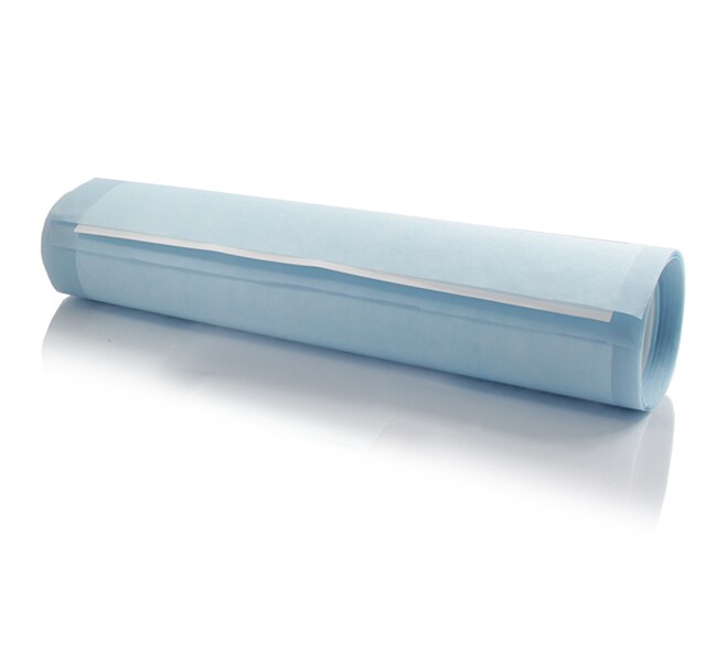
PVDF Transfer Membranes, 0.2 μm, 1 Roll
Pierce PVDF Transfer Membranes are made of high-quality polyvinylidene difluoride and provide high binding capacity for proteins and nucleic acids for western, Southern, and northern blotting methods.
| 货号 | 数量 |
|---|---|
| 88520 | 1 Roll |
货号 88520
价格(CNY)
1,699.00
飞享价
Ends: 27-Jun-2026
8,331.00共减 6,632.00 (80%)
Each
数量:
1 Roll
价格(CNY)
1,699.00
飞享价
Ends: 27-Jun-2026
8,331.00共减 6,632.00 (80%)
Each
Pierce PVDF Transfer Membranes are made of high-quality polyvinylidene difluoride and provide high binding capacity for proteins and nucleic acids for western, Southern, and northern blotting methods. The 0.2-μm pore size is ideal for protein analysis of small amounts of proteins (as little as 10 pmoles), amino acid analysis, and transfer of small molecular weight proteins and peptides. For best results, preactivate the membrane with 100% alcohol.
Features include:
• Re-probe characteristics—high mechanical strength makes PVDF an excellent membrane for stripping and re-probing
• Durability—compatible with most organic solvents, acids, and mild bases
• Compatible with commonly used transfer conditions and detection methods such as staining, chemiluminescence, and radiolabeling. Choose Low-Fluorescence PVDF Transfer Membrane for fluorescent western blotting
• High sensitivity—exceptional binding capacity makes PVDF the ultimate choice for low abundance proteins
For Research Use Only. Not for use in diagnostic procedures.
规格
结合功能Goat IgG: 448 μg/cm2, BSA: 340 μg/cm2, Insulin: 262 μg/cm2
产品类型PVDF Transfer Membrane
数量1 Roll
运输条件Room Temperature
尺寸 (长 x 宽)3.75 m x 26.5 cm
产品规格Roll
长度(公制)3.75 m
材质PVDF
孔径0.2 μm
足够用于≥100 Mini-gel Blots (When cut to 8.8 x 10 cm); ≥80 Midi-gel Blots (When cut to 8.5 x 13.25 cm)
宽度(公制)26.5 cm
Unit SizeEach
内容与储存
Store membranes flat at ambient temperature and away from chemical vapors. Some solvent vapors may partially dissolve the membranes, which will disrupt the pore structure.