Search

Thermo Scientific™
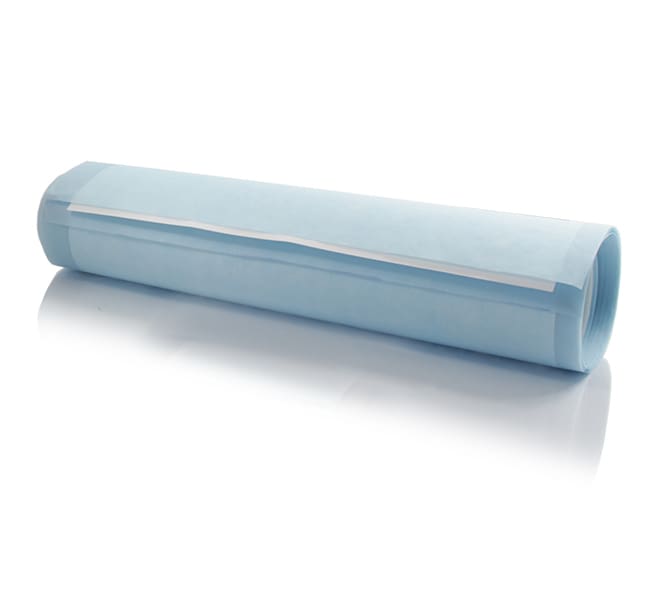
PVDF 转印膜,0.45 μm,10 张预切膜
高质量的聚偏二氟乙烯 (PVDF) 膜,成卷或成片提供,对蛋白质和核酸具有很高的结合能力。
| 货号 | 数量 |
|---|---|
| 88585 | 10 Pre-cut Membranes |
货号 88585
价格(CNY)
2,453.00
Each
数量:
10 Pre-cut Membranes
价格(CNY)
2,453.00
Each
Thermo Scientific™ Pierce PVDF 膜为高质量的聚偏二氟乙烯膜,成卷或成片提供,对蛋白质和核酸具有很高的结合能力,可用于 Western、Southern 和 Northern 印迹法。
这些 PVDF(聚偏二氟乙烯)膜比其他市售 PVDF 膜更耐变色,并且比其他用于 Western 印迹法的转移膜(包括硝酸纤维素)具有更好的吸附蛋白保留能力。标准的 0.45 微米孔径膜有预切割膜片和膜卷可供选择。低荧光 PVDF 膜具有 0.2 微米的孔径,且是与荧光探针配合使用的理想选择。与常规 PVDF 或硝酸纤维素膜相比,该膜的背景水平较低,并且对荧光检测的灵敏度更高。
特点:
Western 印迹法–为 PVDF 转移膜,生产专用于蛋白转移和 Western 印迹的应用
兼容且耐用–聚偏二氟乙烯 (PVDF) 与大多数有机溶剂、酸和温和碱兼容;不会像硝酸纤维素一样破裂或变脆
易于使用–预切割 PVDF 膜片为即用型膜片,可随时用于标准预制和自制微型凝胶 (SDS-PAGE) 的转移和印迹实验
专用选项–可选择 0.45μm 膜,用于比色和化学发光检测;使用低荧光膜 (0.2 μm),用于荧光印迹法
仅供科研使用。不可用于诊断程序。
规格
描述Pierce PVDF 转移膜,0.45um,10cm x 10cm
数量10 Pre-cut Membranes
运输条件Room Temperature
尺寸 (长 x 宽)10 x 10 cm
产品规格片
长度(公制)10 cm
材质PVDF
孔径0.45 μm
足够用于10 Mini-gel Blots
宽度(公制)10 cm
Unit SizeEach
内容与储存
在环境温度下将膜平放储存,远离化学蒸汽。一些溶剂蒸汽可能会溶解部分膜,破坏孔隙结构。