Search


引用和文献 (68)
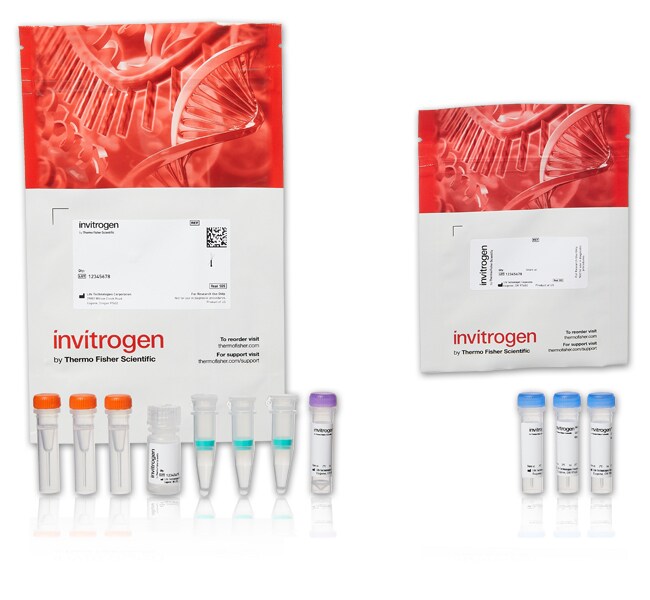
Invitrogen™
Alexa Fluor™ 488 微量蛋白标记试剂盒
微量蛋白标记试剂盒提供了一种便利的方法,可将荧光标记连接至少量抗体或蛋白 (20–100 μg)。试剂盒具有四种 Alexa Fluor™ 颜色(或生物素),提供三次标记和分离反应所需的全部试剂。了解更多信息
| 货号 | 数量 |
|---|---|
| A30006 | 1 个试剂盒 |
货号 A30006
价格(CNY)
10,696.00
Each
数量:
1 个试剂盒
价格(CNY)
10,696.00
Each
微量蛋白标记试剂盒提供了一种便利的方法,可将荧光标记连接至少量抗体或蛋白 (20–100 μg)。试剂盒具有四种 Alexa Fluor™ 颜色(或生物素),提供三次标记和分离反应所需的全部试剂。
微量蛋白标记试剂盒的重要特点:
• 标记蛋白通常可在 2 小时内立即可用(手动操作时间 ∼30 min)
• 针对分子量在 12 和 150 kDa 之间的 20–100 μg 蛋白进行了优化
• 使用便利的离心过滤柱进行纯化,得率介于 60% 和 90% 之间
• 标记前必须去除样品中的稳定蛋白
稳定化学反应和卓越 Alexa Fluor™ 染料
在微量蛋白标记试剂盒中,反应性染料含有四氟苯基 (TFP) 酯部分,该部分在溶液中的稳定性优于常用的琥珀酰亚胺 (NHS) 酯。TFP 酯与蛋白的伯胺高效反应,形成稳定的染料–蛋白偶联物。与传统染料相比,Alexa Fluor™ 染料更明亮、光稳定性更好,且在 pH 值 4 至 10 之间的 pH 值耐受性更强。通常,当使用 Alexa Fluor™ 染料时,可达到更高程度的标记,而无需分子内淬灭。有关详细信息,请参见可见光谱和红外光谱的 Alexa Fluor™染料—第 1.3 节。
了解更多关于蛋白质和抗体标记的信息
我们提供了多种 Molecular Probes™ 抗体和蛋白标记试剂盒以满足您的原材料和实验设置。参见 A 至 Z 的抗体标记或使用我们的标记化学选择工具进行其他选择。了解更多有关我们的各种试剂盒的信息,请参阅 Molecular Probes™ 手册中的蛋白和核酸标记试剂盒—章节 1.2。
我们将为您制备定制的抗体偶联物
如果在我们的库存清单中找不到您需要的产品,我们将为您制备定制的抗体偶联物。我们的定制偶联服务高效且保密, 而且我们绝对保证质量。我们经过ISO 9001:2000认证。
仅供科研使用。不适用于动物或人类的治疗或诊断。
仅供科研使用。不可用于诊断程序。
规格
颜色绿色
检测方法荧光
激发/发射495/519
标签类型Alexa Fluor 染料
标记方法基于偶联
标记规模20 至 100 μg
产品线Alexa Fluor
产品类型蛋白标记试剂盒
数量1 个试剂盒
反应一部分四氟苯基 (TFP) 酯
运输条件室温
标记目标蛋白
标签或染料Alexa Fluor 488
Unit SizeEach
内容与储存
在冷藏冰箱(2°C 至 8°C)中避光储存。
常见问题解答 (FAQ)
我应该使用多大浓度的抗体进行偶联?
何为标记度(DOL)?
Can I use 50 μg of protein with Fluorescent Protein Labeling Kits?
What formulation of antibody should I use for conjugation for small animal in vivo imaging?
What is degree of labeling (DOL)?
引用和文献 (68)
引用和文献
Abstract
Golgi apparatus immunolocalization of endomannosidase suggests post-endoplasmic reticulum glucose trimming: implications for quality control.
Journal:Mol Biol Cell
PubMed ID:11102520
'Trimming of N-linked oligosaccharides by endoplasmic reticulum (ER) glucosidase II is implicated in quality control of protein folding. An alternate glucosidase II-independent deglucosylation pathway exists, in which endo-alpha-mannosidase cleaves internally the glucose-substituted mannose residue of oligosaccharides. By immunogold labeling, we detected most endomannosidase in cis/medial Golgi cisternae (83.8% of immunogold
Glycosylation influences the lectin activities of the macrophage mannose receptor.
Journal:J Biol Chem
PubMed ID:15983039
'The mannose receptor (MR) is a heavily glycosylated endocytic receptor that recognizes both mannosylated and sulfated ligands through its C-type lectin domains and cysteine-rich (CR) domain, respectively. Differential binding properties have been described for MR isolated from different sources, and we hypothesized that this could be due to altered glycosylation.
An endocytosed TGN38 chimeric protein is delivered to the TGN after trafficking through the endocytic recycling compartment in CHO cells.
Journal:J Cell Biol
PubMed ID:9722606
'To examine TGN38 trafficking from the cell surface to the TGN, CHO cells were stably transfected with a chimeric transmembrane protein, TacTGN38. We used fluorescent and 125I-labeled anti-Tac IgG and Fab fragments to follow TacTGN38''s postendocytic trafficking. At steady-state, anti-Tac was mainly in the TGN, but shortly after endocytosis it
Alexa dyes, a series of new fluorescent dyes that yield exceptionally bright, photostable conjugates.
Journal:J Histochem Cytochem
PubMed ID:10449539
'Alexa 350, Alexa 430, Alexa 488, Alexa 532, Alexa 546, Alexa 568, and Alexa 594 dyes are a new series of fluorescent dyes with emission/excitation spectra similar to those of AMCA, Lucifer Yellow, fluorescein, rhodamine 6G, tetramethylrhodamine or Cy3, lissamine rhodamine B, and Texas Red, respectively (the numbers in the
Removal of the membrane-anchoring domain of epidermal growth factor leads to intracrine signaling and disruption of mammary epithelial cell organization.
Journal:J Cell Biol
PubMed ID:9832559
'Autocrine EGF-receptor (EGFR) ligands are normally made as membrane-anchored precursors that are proteolytically processed to yield mature, soluble peptides. To explore the function of the membrane-anchoring domain of EGF, we expressed artificial EGF genes either with or without this structure in human mammary epithelial cells (HMEC). These cells require activation