Search


引用和文献 (5)
Invitrogen™
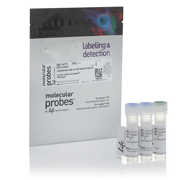
LIVE/DEAD™ 可固定远红外死细胞染色剂试剂盒,用于 633 或 635 nm 激发
在细胞内抗体染色所需的固定和透化前,或者在使用甲醛固定消除生物危害性物质前,使用 LIVE/DEAD™ 可固定远红外死细胞染色剂试剂盒测定细胞活力。本试剂盒经过优化和验证,可与红色激光流式细胞仪配合使用。•稳定—染料在单独小瓶中冷冻干燥,以维持稳定性• 稳健—染色模式在固定前后相同• 明亮信号—能够以单通道轻松分辨活/死细胞查看用于流式细胞分析的所有可固定活力染料的选择指南。稳定与以溶液形式销售的产品不同,LIVE/DEAD™ 可固定远红外染色剂以 40了解更多信息
Have Questions?
更改视图

| 货号 | 数量 |
|---|---|
| L34973 | 80 次检测 |
| L34974 | 400 次检测 |
| L10120 | 200 次检测 |
货号 L34973
价格(CNY)
1,131.00
飞享价
Ends: 31-Dec-2026
1,503.00共减 372.00 (25%)
Each
数量:
80 次检测
价格(CNY)
1,131.00
飞享价
Ends: 31-Dec-2026
1,503.00共减 372.00 (25%)
Each
在细胞内抗体染色所需的固定和透化前,或者在使用甲醛固定消除生物危害性物质前,使用 LIVE/DEAD™ 可固定远红外死细胞染色剂试剂盒测定细胞活力。本试剂盒经过优化和验证,可与红色激光流式细胞仪配合使用。
•稳定—染料在单独小瓶中冷冻干燥,以维持稳定性
• 稳健—染色模式在固定前后相同
• 明亮信号—能够以单通道轻松分辨活/死细胞
查看用于流式细胞分析的所有可固定活力染料的选择指南。
稳定
与以溶液形式销售的产品不同,LIVE/DEAD™ 可固定远红外染色剂以 40 次测试瓶便利包装,有助于确保染料随时间推移的稳定性和性能。溶液中的胺反应性染料在短时间内会失去其有效性,因此建议在再水化后完全使用小瓶。如果无法做到这一点,将小瓶以小体积等分,并储存在 -80°C 下,避免冻融循环。
稳健
死细胞鉴别染色剂可在固定剂(如细胞内磷酸化研究所需的甲醛或基于乙醇的固定方法)处理后失去敏感性。LIVE/DEAD™ 可固定远红外染色剂是一种与细胞内和细胞外胺共价结合的胺反应性染料,在甲醛固定后保留染色模式。
极佳亮度
LIVE/DEAD™ 可固定远红外染色剂根据其在使用红色激光激发时提供明亮信号的荧光特性所选择。远红外荧光反应性染料的最大激发波长为 ∼633 nm,使其适用于与发射波长为 ∼655 nm 的红色或 HeNe 激光配合使用。因为可以使用流式细胞仪的单色染料和单通道来区分活细胞和死细胞、所以它是多色实验的理想选择。
工作原理
在细胞膜受损的细胞中,染料可与细胞内部和细胞表面的游离胺基反应,产生强烈的荧光染色。在活细胞中,染料的反应活性仅限于细胞表面的胺,从而导致更少的荧光强度。活细胞与死细胞之间的强度差异通常大于 50 倍,可轻松分辨。
提供多种颜色
LIVE/DEAD™ 可固定死细胞染色剂有多种颜色可供选择,以满足您的多色检测组合需求。
仅供科研使用。不可用于诊断程序。
规格
细胞渗透性不透过
细胞类型真核细胞
描述LIVE/DEAD™ 可固定远红外死细胞染色剂试剂盒,用于 633 或 635 nm 激发
检测方法荧光
染料类型LIVE/DEAD™ 可固定远红外死细胞染色剂
形式实心
产品规格管
数量80 次检测
运输条件室温
溶解度DMSO(二甲亚砜)
颜色远红光
发射655 nm
Excitation Wavelength Range633 nm
适用于(应用)活力测定试剂盒
适用于(设备)流式细胞仪
产品线LIVE/DEAD
产品类型染色剂
Unit SizeEach
内容与储存
包含2小瓶 LIVE/DEAD™ 可固定死细胞染色剂以及 500 μL DMSO。
在 -20°C 下储存。
在 -20°C 下储存。
常见问题解答 (FAQ)
我如何为LIVE/DEAD细胞活力实验准备死细胞对照?
哪一种细胞活力试剂盒兼容固定?
How do I prepare dead cell controls for LIVE/DEAD cell viability assays?
Which cell viability kits are compatible with fixation?
引用和文献 (5)
引用和文献
Abstract
Levels of circulating endothelial cells are low in idiopathic pulmonary fibrosis and are further reduced by anti-fibrotic treatments.
Journal:
PubMed ID:26552487
'It has been suggested that circulating fibrocytes and endothelial cells actively participate in the intense remodelling of the pulmonary vasculature in patients with idiopathic pulmonary fibrosis (IPF). Indeed, fibrotic areas exist that have fewer blood vessels, whereas adjacent non-fibrotic tissue is highly vascularized. The number of circulating endothelial cells (CEC)
The oxysterol-CXCR2 axis plays a key role in the recruitment of tumor-promoting neutrophils.
Journal:
PubMed ID:23897983
'Tumor-infiltrating immune cells can be conditioned by molecules released within the microenvironment to thwart antitumor immune responses, thereby facilitating tumor growth. Among immune cells, neutrophils play an important protumorigenic role by favoring neoangiogenesis and/or by suppressing antitumor immune responses. Tumor-derived oxysterols have recently been shown to favor tumor growth by
CD8+ T cell responses to lytic EBV infection: late antigen specificities as subdominant components of the total response.
Journal:
PubMed ID:24146041
EBV elicits primary CD8(+) T cell responses that, by T cell cloning from infectious mononucleosis (IM) patients, appear skewed toward immediate early (IE) and some early (E) lytic cycle proteins, with late (L) proteins rarely targeted. However, L Ag-specific responses have been detected regularly in polyclonal T cell cultures from
Deficiency of terminal ADP-ribose protein glycohydrolase TARG1/C6orf130 in neurodegenerative disease.
Journal:EMBO J
PubMed ID:23481255
Adenosine diphosphate (ADP)-ribosylation is a post-translational protein modification implicated in the regulation of a range of cellular processes. A family of proteins that catalyse ADP-ribosylation reactions are the poly(ADP-ribose) (PAR) polymerases (PARPs). PARPs covalently attach an ADP-ribose nucleotide to target proteins and some PARP family members can subsequently add additional
G-CSF activation of AKT is not sufficient to prolong neutrophil survival.
Journal:J Leukoc Biol
PubMed ID:23559492
Neutrophils play an important role in the innate immune response against bacterial and fungal infections. They have a short lifespan in circulation, and their survival can be modulated by several cytokines, including G-CSF. Previous studies have implicated AKT as a critical signaling intermediary in the regulation of neutrophil survival. Our