流式细胞仪能分析哪些细胞过程?
•钙流:每一种Oregon Green钙指示剂都可通过更高的亲和力结合胞内钙离子,提供适合很多应用的灵敏度范围。Oregon Green探针在Ca2+静息水平下发射绿色荧光;Ca2+浓度增加时,荧光强度会增加14倍。细胞通透配方(货号O6807)能加入到细胞培养基中并与流式细胞仪兼容。
•基于罗丹明的钙离子指示剂包含了大量不同的探针,用于探测Ca2+浓度的大小变化。该指示剂与钙离子结合后,会发出50倍增强的荧光。这一类波长范围的荧光可与GFP或绿色荧光染料结合,用于多重检测应用。Rhod-2, AM(货号R1245MP)专门靶向线粒体,可与流式细胞仪联用。
•膜电位:细胞凋亡初期的典型特征是线粒体紊乱,伴随着膜和氧化还原电位变化。我们提供一系列专门用于流式细胞术活细胞线粒体膜电位分析的产品,可最大限度避免影响细胞功能。对于细胞凋亡过程出现的线粒体膜电位损失,MitoProbe系列线粒体染色剂(货号M34150、M34151和M34152)可提供快速、简单和可靠的流式细胞术检测方法。MitoTracker染料(货号M7510和M7512)是用于染色活细胞内的线粒体的膜电位依赖型探针。在之后的流式细胞免疫化学、DNA末端标记,原位杂交或复染色步骤中,MitoTracker染料的染色图案全程保留。相比于只依赖线粒体膜电位的检测方法,线粒体通透性转移孔检测体系(货号M34153)可直接测定通透性转移孔开合情况。线粒体通透性转移孔(MPTP)是一个由线粒体内膜和外膜成分构成的非特异性通道。在细胞死亡过程中,此通道显现,参与线粒体成分释放。
•吞噬作用:在吞噬过程中,细胞内吞微粒(如微生物)。此过程对于免疫应答非常重要,同时对清除凋亡细胞也非常重要。研究吞噬作用的探针包括BioParticles指示剂——用荧光标记的细菌和酵母。
•使用淬灭/洗涤检测法来追踪吞噬过程能够表征简单的摄入,或利用一种pH指示剂监视吞噬途径的各个阶段。我们提供pHrodo Red或Green(货号A10010、P35361、P35364、P35365、P35366和P35367)标记的免洗检测体系和全血的免洗检测体系(货号A10025、A10026、P35381和P35382),都适用于流式细胞仪。
•pH改变:可使用荧光强度或比率计测定法测定生理学范围内的细微pH变化。pHrodo 染料(货号P35373和P35372)提供了pH2-9之间的信号强度调制,同时可以选择各种荧光波长。荧光右旋糖酐内吞示踪是分析细胞区室pH变化的常用方法。pHrodo染料的右旋糖酐偶联物(货号P35368和P10361)可用于区分从早期的核内体到溶酶体在内的各种囊泡,不需要洗涤和淬灭,是最完整的解决方案。
•活性氧:处于环境压力下的细胞通常含有超高水平的活性氧(ROS)。CellROX试剂是为检测和定量活细胞中的ROS而开发的荧光探针。这些细胞通透性试剂在还原态时不发荧光或发微弱的荧光;在被氧化后,就发出明亮的荧光且依旧位于细胞内。我们提供已通过流式细胞术验证的CellROX Green(货号C10492)CellROX Orange(货号C10493)和CellROX Deep Red(货号C10491)检测试剂盒。
Can I store reconstituted pHrodo BioParticles Conjugates for Phagocytosis and Phagocytosis Kit, for Flow Cytometry?
Yes. Once reconstituted, pHrodo BioParticles Conjugates for Phagocytosis and Phagocytosis Kit, for Flow Cytometry (Cat. Nos. P35367, P35361, P35360, P35366, P35364, P35365, A10010) can be stored at 2 - 8 degrees C for several weeks, as long as sodium azide is added to a final concentration of 2 mM. If no sodium azide is added, the cell suspension needs to be used right away or on the same day to avoid contamination. DO NOT FREEZE the resuspended pHrodo bioparticle conjugates.
Find additional tips, troubleshooting help, and resources within our Cell Analysis Support Center.
Are the Invitrogen BioParticles products sterile?
While the bacteria have been attenuated with formaldehyde and alcohol desiccation, the BioParticles products are not considered sterile, and we do not recommend incubation of more than 4 hours. This applies to all of our dye-labeled (pHrodo, Alexa Fluor, etc.) and unlabeled BioParticles products.
What is the type of bond that attaches the dyes to the BioParticles probes?
We use amine-reactive dyes to covalently attach fluorescent dyes to all of our BioParticles probes such as the Escherichia coli (K-12 strain) BioParticles probes, Staphylococcus aureus (Wood strain without protein A) BioParticles, and the Zymosan A (S. cerevisiae) BioParticles probes.
Find additional tips, troubleshooting help, and resources within our Cell Analysis Support Center.
What cellular processes can be analyzed with a flow cytometer?
-Calcium flux: Each of the Oregon Green calcium indicators binds intracellular calcium with increasing affinity, providing a sensitivity range to match many applications. Oregon Green probes emit green fluorescence at resting levels of Ca2+ and increase their fluorescence intensity 14-fold with increasing Ca2+ concentration. The cell-permeant formulation (Cat. No. O6807) can be loaded in cell media and is compatible with flow cytometry.
-Rhodamine-based calcium indicators comprise a range of probes for large or small changes in Ca2+ concentration. They exhibit a 50-fold increase in fluorescence upon calcium binding and offer a range of wavelengths that can be used in conjunction with GFP or green-fluorescent dyes for multiplexing. Rhod-2, AM (Cat. No. R1245MP), in particular, localizes to mitochondria and can be used with flow cytometry.
-Membrane potential: A distinctive feature of the early stages of apoptosis is the disruption of the mitochondria, including changes in membrane and redox potential. We offer a range of products specifically designed to assay mitochondrial membrane potential in live cells by flow cytometry, with minimal disruption of cellular function. The MitoProbe family of mitochondrial stains (Cat. Nos. M34150, M34151, and M34152) provide quick, easy, and reliable flow cytometric detection of the loss of mitochondrial membrane potential that occurs during apoptosis. MitoTracker dyes (Cat. Nos. M7510 and M7512) are membrane potential-dependent probes for staining mitochondria in live cells. The staining pattern of MitoTracker dyes is retained throughout subsequent flow cytometry immunocytochemistry, DNA end labeling, in situ hybridization, or counterstaining steps. The Mitochondrial Permeability Transition Pore Assay (Cat. No. M34153) provides a more direct method of measuring mitochondrial permeability transition pore opening than assays relying on mitochondrial membrane potential alone. The mitochondrial permeability transition pore (MPTP) is a non-specific channel formed by components from the inner and outer mitochondrial membranes, and appears to be involved in the release of mitochondrial components during cell death.
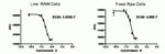
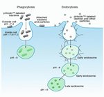
-Phagocytosis: In phagocytosis, cells internalize particulate matter such as microorganisms, and this process is important for immune responses and during the clearance of apoptotic cells. Probes for studying phagocytosis include BioParticles indicators—bacteria and yeast labeled with fluorescent dyes.
-Tracking phagocytosis using a quench/wash-based assay can report on simple uptake, or a pH indicator can be used to monitor stages in the pathway. We have no-wash assays labeled with pHrodo Red or Green (Cat. Nos. A10010, P35361, P35364, P35365, P35366, and P35367) and no-wash assays for whole blood (Cat. Nos. A10025, A10026, P35381, and P35382), all suitable for flow cytometry.
-pH changes: Sensitive pH determinations can be made in a physiological range using either fluorescent intensity or ratiometric measurements. pHrodo dyes (Cat. Nos. P35373 and P35372) provide signal intensity modulation from pH 2 to pH 9 and with a choice of fluorescent wavelengths. Tracking internalization of fluorescent dextran is a routine method for analyzing pH changes in cellular compartments. Dextran conjugates of pHrodo dyes (Cat. Nos. P35368 and P10361) provide the most complete solution by allowing discrimination of vesicles from early endosomes to lysosomes, with no quench or wash required.
-Reactive oxygen species: Cells that are environmentally stressed usually contain greatly increased levels of reactive oxygen species (ROS). CellROX reagents are fluorogenic probes developed for the detection and quantitation of ROS in live cells. These cell-permeant reagents are non-fluorescent or very weakly fluorescent in the reduced state; however, when oxidized, they become brightly fluorescent and remain localized within the cell. We offer CellROX Green (Cat. No. C10492), CellROX Orange (Cat. No. C10493), and CellROX Deep Red (Cat. No. C10491) Assay Kits validated for flow cytometry.
Find additional tips, troubleshooting help, and resources within our Cell Analysis Support Center.