Search
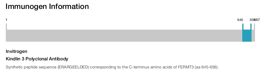
Invitrogen
Kindlin 3 Polyclonal Antibody
{{$productOrderCtrl.translations['antibody.pdp.commerceCard.promotion.promotions']}}
{{$productOrderCtrl.translations['antibody.pdp.commerceCard.promotion.viewpromo']}}
{{$productOrderCtrl.translations['antibody.pdp.commerceCard.promotion.promocode']}}: {{promo.promoCode}} {{promo.promoTitle}} {{promo.promoDescription}}. {{$productOrderCtrl.translations['antibody.pdp.commerceCard.promotion.learnmore']}}
Additional Information:
{{banner.description}}
")
图: 1 / 1
Kindlin 3 Antibody (PA5-19174) in WB
Western blot analysis of Kindlin 3 using Kindlin 3 Polyclonal Antibody (Product # PA5-19174) (1 µg/mL) in staining of Human Peripheral Blood Mononucleocytes lysate (35 µg protein in RIPA buffer). Primary incubation was 1 hour. Detected by chemiluminescence.
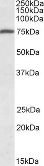
Please note: We are reviewing Western blot images included in the antibody testing data in our catalog, including those provided by third parties. Unless expressly labeled or annotated as “raw-unedited”, Western blot images included in the antibody testing data in our catalog may have been edited, optimized or otherwise adjusted for presentation.
 in WB")
产品信息
PA5-19174
产品规格
种属反应
Human
宿主/亚型
Goat
/ IgG
分类
Polyclonal
类型
Antibody
抗原
Synthetic peptide sequence (ERARGEELDED) corresponding to the C-terminus amino acids of FERMT3 (aa 645-656).
偶联物
Unconjugated
Unconjugated
Unconjugated
形式
Liquid
浓度
0.5 mg/mL
规格
100 µg
纯化类型
Ammonium sulfate precipitation
保存液
TBS, pH 7.3, with 0.5% BSA
内含物
0.02% sodium azide
保存条件
-20°C, Avoid Freeze/Thaw Cycles
运输条件
Ambient (domestic); Wet ice (international)
RRID
产品详细信息
This antibody is predicted to react with canine, mouse and rat based on sequence homology.
This antibody is tested in Peptide ELISA: antibody detection limit dilution 2,000.
靶标信息
The three KINDLINs are a novel family of focal adhesion proteins, localizing to integrin adhesion sites. The KINDLIN proteins are composed of a centrally located FERM domain interrupted by a pleckstrin homology (PH) domain. KINDLIN1 and KINDLIN2 have been shown to play an essential role in integrin-mediated adhesion and spreading. In contrast to the widely expressed KINDLIN1 and KINDLIN2, KINDLIN3 is restricted to hematopoietic cells and is particularly abundant in megakaryocytes and platelets. Several reports describe a transcriptional misregulation of KINDLINs in various types of cancer. A recent study demonstrates that KINDLIN3 is essential for platelet integrin activation and subsequent integrin outside-in signaling, suggesting it may serve as a potential target for the design of therapeutics aimed at specifically disrupting integrin activation in platelets and leukocytes.
仅用于科研。不用于诊断过程。未经明确授权不得转售。
篇参考文献 (0)
您是否在文献中引用过该产品?请点击下方按钮邮件告知我们。
生物信息学
蛋白别名: MGC10966
Disclaimer

Clicking the images or links will redirect you to a website hosted by BenchSci that provides third-party scientific content. Neither the content nor the BenchSci technology and processes for selection have been evaluated by us; we are providing them as-is and without warranty of any kind, including for use or application of the Thermo Fisher Scientific products presented.

