Search


引用和文献 (15)
Invitrogen™
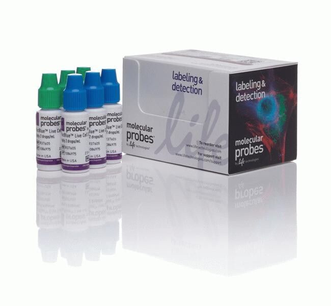
ReadyProbes™ 细胞活力成像试剂盒(蓝色/绿色)
ReadyProbes® 细胞活力成像试剂盒(蓝色/绿色)是一种即用型检测,可快速且轻松地测定细胞活力。只需向 1 mL 细胞生长培养基中加入室温下的稳定 NucBlue® Live了解更多信息
| 货号 | 数量 |
|---|---|
| R37609 | 1 kit |
货号 R37609
价格(CNY)
2,460.00
飞享价
Ends: 31-Dec-2026
3,416.00共减 956.00 (28%)
Each
数量:
1 kit
价格(CNY)
2,460.00
飞享价
Ends: 31-Dec-2026
3,416.00共减 956.00 (28%)
Each
ReadyProbes® 细胞活力成像试剂盒(蓝色/绿色)是一种即用型检测,可快速且轻松地测定细胞活力。只需向 1 mL 细胞生长培养基中加入室温下的稳定 NucBlue® Live 试剂 (Hoechst 33342) 和 NucGreen® Dead 试剂各2滴,然后即可通过计数总细胞与死细胞来确定细胞活力。NucBlue® Live 试剂可对所有细胞的细胞核进行染色,并可使用标准 DAPI 滤光片进行检测。NucGreen® Dead 试剂仅对细胞膜完整性受损细胞的细胞核进行染色,并能使用标准 FITC/GFP(绿色)滤光片组进行检测。本试剂盒适用于荧光显微镜、荧光微孔板读数仪和流式细胞分析。
NucBlue® Live 试剂: 可对所有细胞的细胞核进行染色;使用标准 DAPI 滤光片进行检测(最大激发/发射波长:360/460 nm)
NucGreen® Dead 试剂: 仅对细胞膜受损的死细胞的细胞核进行染色;使用标准 FITC/GFP(绿色)滤光片组进行检测(最大激发/发射波长:504/523 nm)
请参阅其他 ReadyProbes® 细胞染色试剂
进一步了解其他细胞活力测定试剂盒
使用建议
•NucBlue® Live 和 NucGreen® Dead 试剂可直接加入完全生长培养基的细胞或兼容的缓冲液中。
• 在大多数情况下,2滴/mL 以及5至30分钟的孵育时间将使细胞核染上明亮颜色;然而,可能需要针对一些细胞类型、条件和应用进行优化。在此种情况下,只需简单地加入更多或更少的液滴,直至获得较佳染色强度。
仅供科研使用。不可用于诊断程序。
规格
细胞类型哺乳动物细胞、真核细胞
描述ReadyProbes 细胞活力成像试剂盒,蓝色/绿色
检测方法荧光
染料类型3 x 2.5 mL NucBlue™ Live
产品规格滴瓶
数量1 kit
运输条件室温
颜色蓝色、绿色
发射360、504 nm
Excitation Wavelength Range460、523 nm
适用于(应用)活力测定试剂盒
适用于(设备)荧光显微镜
产品线ReadyProbes
产品类型细胞活力成像试剂盒
Unit SizeEach
内容与储存
在 2° 至 8°C 或室温下储存。
常见问题解答 (FAQ)
我在 Countess II FL全自动细胞计数仪上能进行哪种荧光活性实验?
Can I use the ReadyProbes reagents for flow cytometry?
What fluorescent viability assays can I use on the Countess II FL automated cell counter?
What is the difference between NucBlue Live reagents stain and NucGreen Dead reagents stain?
引用和文献 (15)
引用和文献
Abstract
In vitro studies with renal proximal tubule cells show direct cytotoxicity of Androctonus australis hector scorpion venom triggered by oxidative stress, caspase activation and apoptosis.
Journal:Toxicon
PubMed ID:27470530
'Scorpion envenomation injures a number of organs, including the kidney. Mechanisms proposed to explain the renal tubule injury include direct effects of venom on tubule epithelial cells, as well as indirect effects of the autonomic nervous system, and inflammation. Here, we report direct effects of Androctonus australis hector (Aah) scorpion
Fluorescence and Cytotoxicity of Cadmium Sulfide Quantum Dots Stabilized on Clay Nanotubes.
Journal:Nanomaterials (Basel)
PubMed ID:29857546
'Quantum dots (QD) are widely used for cellular labeling due to enhanced brightness, resistance to photobleaching, and multicolor light emissions. CdS and Cd'
Chemobrionic structures in tissue engineering: self-assembling calcium phosphate tubes as cellular scaffolds.
Journal:Biomater Sci
PubMed ID:31830151
'A diverse range of complex patterns and mineralised hierarchical microstructures can be derived from chemobrionic systems, with formation driven by complex reaction-diffusion mechanisms far from thermodynamic equilibrium. In these experiments, self-assembling calcium phosphate tubes are generated using hydrogels made with 1 M calcium solutions layered with solutions of dibasic sodium
An evolutionary perspective on the role of mesencephalic astrocyte-derived neurotrophic factor (MANF): At the crossroads of poriferan innate immune and apoptotic pathways.
Journal:Biochem Biophys Rep
PubMed ID:28955781
'The mesencephalic astrocyte-derived neurotrophic factor (MANF) belongs to a recently discovered family of neurotrophic factors. MANF can be secreted but is generally resident within the endoplasmic reticulum (ER) in neuronal and non-neuronal cells, where it is involved in the ER stress response with pro-survival effects. Here we report the discovery
A novel method to understand tumor cell invasion: integrating extracellular matrix mimicking layers in microfluidic chips by
Journal:Biomed Microdevices
PubMed ID:29038872
'A major challenge in studying tumor cell invasion into its surrounding tissue is to identify the contribution of individual factors in the tumor microenvironment (TME) to the process. One of the important elements of the TME is the fibrous extracellular matrix (ECM) which is known to influence cancer cell invasion,