Search


引用和文献 (20)
Invitrogen™
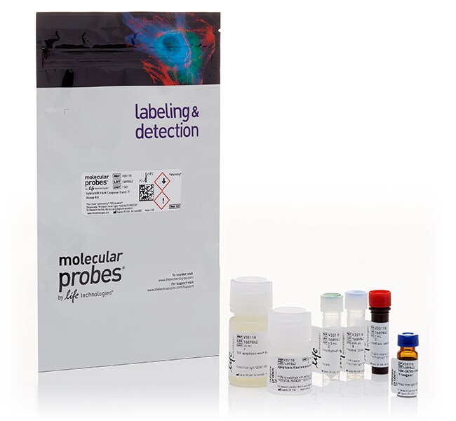
用于流式细胞分析的 Vybrant™ FLICA 半胱天冬酶细胞凋亡检测试剂盒
采用用于检测多聚半胱天冬酶和半胱天冬酶-3/7/8 活性的 Vybrant FLICA 半胱天冬酶检测试剂盒,通过流式细胞分析检测细胞凋亡情况。
Have Questions?
更改视图

| 货号 | 酶 |
|---|---|
| V35118 | Caspase 3/7 |
| V35117 | Poly Caspases |
| V35119 | Caspase 8 |
货号 V35118
价格(CNY)
3,556.00
飞享价
Ends: 31-Dec-2026
4,723.00共减 1,167.00 (25%)
25 assays
酶:
Caspase 3/7
价格(CNY)
3,556.00
飞享价
Ends: 31-Dec-2026
4,723.00共减 1,167.00 (25%)
25 assays
使用适用于多聚半胱天冬酶以及半胱天冬酶 3 和 7 的 Vybrant FAM 半胱天冬酶检测试剂盒,通过流式细胞分析,快速检测半胱天冬酶介导的细胞凋亡。Vybrant FAM 半胱天冬酶检测试剂盒使用荧光半胱天冬酶抑制剂 (FLICA) 方法,用于检测和报告半胱天冬酶活性。具体而言,这些半胱天冬酶检测试剂盒包含经工程改造的 FLICA 底物 FAM-VAD-FMK(用于多聚半胱天冬酶)、FAM-DEVD-FMK(用于半胱天冬酶-3/7)和 FAM-LETD-FMK(用于半胱天冬酶-8)。它们还包括 Hoechst 33342 和碘化丙啶染色剂,能够通过流式细胞分析同时评估膜可透过性和细胞周期。
用于流式细胞分析的 Vybrant FAM 半胱天冬酶检测试剂盒采用了一种检测活性半胱天冬酶的新方法:该试剂盒利用 FLICA 方法(亲和标记)。该亲和标记将一个可以与半胱氨酸发生共价反应的氟甲基酮 (FMK) 部分和一个半胱天冬酶特异性氨基酸序列相连接。不同的氨基酸识别序列用于检测不同的半胱天冬酶:缬氨酸-丙氨酸-天冬氨酸 (VAD) 用于检测多聚半胱天冬酶(包括半胱天冬酶-1、-3、-4、-5、-6、-7、-8 和 -9),天冬氨酸-谷氨酸-缬氨酸-天冬氨酸 (DEVD) 用于检测半胱天冬酶-3/7,亮氨酸-谷氨酸-苏氨酸-天冬氨酸 (LETD) 用于检测半胱天冬酶-8。连有一个羧基荧光素 (FAM) 基团,作为报告基团。
FLICA 试剂与已激活半胱天冬酶的反应中心通过识别序列相互作用,之后通过 FMK 基团与半胱氨酸共价连接。FLICA 抑制剂具有细胞渗透性但没有细胞毒性。未结合的 FLICA 分子扩散出细胞并被洗涤掉,其余的荧光信号可用于直接测定加入抑制剂时存在的活性半胱天冬酶的量。FLICA、碘化丙啶和 Hoechst 33342 试剂的近似激发峰和发射峰分别为 488 nm/⁄530 nm、535⁄ nm/6617 nm 和 350 nm/⁄461 nm。Vybrant FAM 多聚半胱天冬酶检测、半胱天冬酶-3 和半胱天冬酶-7 检测以及半胱天冬酶-8 检测细胞凋亡试剂盒中包括的检测试剂分别为 488⁄⁄530 FAM-VAD-FMK 试剂、488/⁄530 FAM-DEVD-FMK 和 488/⁄530 FAM-LETD-FMK。
FLICA 试剂与已激活半胱天冬酶的反应中心通过识别序列相互作用,之后通过 FMK 基团与半胱氨酸共价连接。FLICA 抑制剂具有细胞渗透性但没有细胞毒性。未结合的 FLICA 分子扩散出细胞并被洗涤掉,其余的荧光信号可用于直接测定加入抑制剂时存在的活性半胱天冬酶的量。FLICA、碘化丙啶和 Hoechst 33342 试剂的近似激发峰和发射峰分别为 488 nm/⁄530 nm、535⁄ nm/6617 nm 和 350 nm/⁄461 nm。Vybrant FAM 多聚半胱天冬酶检测、半胱天冬酶-3 和半胱天冬酶-7 检测以及半胱天冬酶-8 检测细胞凋亡试剂盒中包括的检测试剂分别为 488⁄⁄530 FAM-VAD-FMK 试剂、488/⁄530 FAM-DEVD-FMK 和 488/⁄530 FAM-LETD-FMK。
仅供科研使用。不可用于诊断程序。
规格
酶Caspase 3/7
激发/发射FAM-DEVD-FMD:388/530 PI:535/617 Hoechst 33342:350/461
流式细胞仪激光线路UV,488
适用于(设备)流式细胞仪
标签类型其他标记或染料
标签或染料FAM、Hoechst 33342、碘化丙啶
产品线Vybrant
产品类型半胱天冬酶检测试剂盒
数量25 Assays
运输条件室温
检测方法荧光
产品规格管装
Unit Size25 assays
内容与储存
1 小瓶 FAM-DEVD-FMK 半胱天冬酶-3 和半胱天冬酶-7 试剂(FLICA 试剂、冻干固体)、1 小瓶 Hoechst 33342(400 μL,1 mM 水溶液)、1 小瓶 PI(1 mL,250 μg/mL 水溶液)、1 小瓶 DMSO (0.5 mL)、1 瓶细胞凋亡固定剂溶液(10% 甲醛甲醇 PBS 溶液,6 mL)和 1 瓶 10x 细胞凋亡洗涤缓冲液。避光储存在冷藏冰箱 (2-8°C) 中。
常见问题解答 (FAQ)
What are the fluorescence excitation/emission maxima for the FAM-DEVD-FMK FLICA reagent, Hoechst 33342, and propidium iodide supplied in the Vybrant FAM Caspase-3 and -7 Assay Kit, for flow cytometry?
引用和文献 (20)
引用和文献
Abstract
Monocyte Chemoattractant Protein-Induced Protein 1 (MCPIP1) Enhances Angiogenic and Cardiomyogenic Potential of Murine Bone Marrow-Derived Mesenchymal Stem Cells.
Journal:
PubMed ID:26214508
'The current evidence suggests that beneficial effects of mesenchymal stem cells (MSCs) toward myocardial repair are largely due to paracrine actions of several factors. Although Monocyte chemoattractant protein-induced protein 1 (MCPIP1) is involved in the regulation of inflammatory response, apoptosis and angiogenesis, whether MCPIP1 plays any role in stem cell-induced
Multiparametric evaluation of apoptosis: effects of standard cytotoxic agents and the cyanoguanidine CHS 828.
Journal:Mol Cancer Ther
PubMed ID:15141009
'A multiparametric high-content screening assay for measurement of apoptosis was developed. HeLa cells and lymphoma U-937 cells were exposed to cytotoxic drugs in flat-bottomed optical microtiter plates. After incubation, the DNA-binding dye Hoechst 33342, fluorescein-tagged probes that covalently bind active caspases and chloromethyl-X-rosamine to detect mitochondrial membrane potential (MMP) were
Flow cytometry-based apoptosis detection.
Journal:Methods Mol Biol
PubMed ID:19609746
'An apoptosing cell demonstrates multitude of characteristic morphological and biochemical features, which vary depending on the stimuli and the cell type. The gross majority of classical apoptotic hallmarks can be rapidly examined by flow and image cytometry. Cytometry thus became a technology of choice in diverse studies of cellular demise.
BH3-only protein Noxa regulates apoptosis in activated B cells and controls high-affinity antibody formation.
Journal:Blood
PubMed ID:22144184
The efficiency of humoral immune responses depends on the selective outgrowth of B cells and plasma cells that produce high affinity antibodies. The factors responsible for affinity maturation of B cell clones in the germinal center (GC) have been well established but selection mechanisms that allow clones to enter the
In vitro primary human lymphocyte flow cytometry based micronucleus assay: simultaneous assessment of cell proliferation, apoptosis and MN frequency.
Journal:Mutagenesis
PubMed ID:21791709
In order to minimise the number of positive in vitro cytogenetic results which are not confirmed in rodent carcinogenicity tests, biological systems that are p53 and DNA repair proficient should be recommended. Moreover, an appropriate cytotoxicity parameter for top dose selection should be considered. Recent International Conference on Harmonisation draft