Search


引用和文献 (30)
Invitrogen™
用于细胞凋亡检测的 Annexin V 偶联物
采用流式细胞分析利用 Annexin V 独立 Alexa Fluor、APC、Pacific Blue、PE、FITC 和生物素偶联物检测早期细胞凋亡。
Have Questions?
更改视图

| 货号 | 激发/发射 | 流式细胞仪激光线路 | 偶联物 |
|---|---|---|---|
| A23204 | 650/665 | 633-637 | Alexa Fluor 647 |
| A13201 | 495/519 | 488 | Alexa Fluor 488 |
| A13199 | 494/518 | 488 | FITC |
| A13202 | 578/603 | 532、561 | Alexa Fluor 568 |
| A13203 | 590/617 | 532 | Alexa Fluor 594 |
| A13204 | 生物素-X | ||
| A23202 | 346/442 | UV | Alexa Fluor 350 |
| A35108 | 555/565 | 532、561 | Alexa Fluor 555 |
| A35109 | 679/702 | 633-637 | Alexa Fluor 680 |
| A35110 | 650/660 | 633-637 | APC(别藻蓝素) |
| A35111 | 565/578 | 488, 532, 561 | PE |
| A35122 | 410/455 | 405 | Pacific Blue |
货号 A23204
价格(CNY)
3,702.00
Online Exclusive
Ends: 31-Dec-2026
4,918.00共减 1,216.00 (25%)
Each
激发/发射:
650/665
流式细胞仪激光线路:
633-637
偶联物:
Alexa Fluor 647
价格(CNY)
3,702.00
Online Exclusive
Ends: 31-Dec-2026
4,918.00共减 1,216.00 (25%)
Each
采用用于细胞凋亡检测的 Annexin V 独立偶联物,可快速而可靠地检测早期细胞凋亡。使用流式细胞分析,Annexin V 偶联物在凋亡细胞与非凋亡细胞之间提供达 100 倍的荧光信号强度差异。
膜联蛋白 V 对磷脂酰丝氨酸 (PS) 具有高亲和力,PS 可暴露于正在凋亡的细胞的外叶上。由于具有这种亲和力,荧光标记的膜联蛋白 V 试剂是细胞凋亡研究中常用的试剂。
Annexin V 偶联物为研究磷脂酰丝氨酸(是细胞凋亡中期标志)的外化提供了快速而可靠的检测方法。经我们的荧光膜联蛋白V偶联物染色后,凋亡细胞和未凋亡细胞显示出不同的荧光强度,流式细胞分析它们的差异通常约为 100 倍。
与 Nexins Research BV 合作,我们提供了较佳且较亮的膜联蛋白 V 偶联物,包括 Alexa Fluor 350、488、555、568、594、647 和 680 膜联蛋白 V 偶联物以及 Annexin V APC、生物素-X、FITC、Pacific Blue 和 PE 偶联物。高荧光性膜联蛋白 V 偶联物为研究磷酯酰丝氨酸的外翻提供了快速而可靠的检测方法,它是细胞凋亡早期指标之一。
Annexin V Pacific Blue 偶联物具有紫色可激发性,这使其成为紫色激光仪器和包括绿色或红色荧光染料的多色实验的理想选择。
我们的膜联蛋白V偶联物的优势包括:
• 与 Invitrogen Alexa Fluor 和 eFluor 染料偶联可产生较亮信号
• 偶联物适用于所有可用激光
• 可用作独立试剂或易于使用的试剂盒
Annexin V 染色检测凋亡细胞仅可在活细胞和组织上进行。如果样品染色后被固定,则需要特定的条件来保留信号。包括使用不含酒精的乙醛固定方法、使用含 Ca2+ 但不含表面活性剂/去污剂的缓冲液。为方便您使用,我们还提供了浓缩膜联蛋白结合缓冲液,用于在细胞凋亡检测中促进膜联蛋白 V 与磷脂酰丝氨酸结合。
仅供科研使用。不可用于诊断程序。
规格
颜色远红外
描述Annexin V Alexa Fluor 647 偶联物
激发/发射650/665
流式细胞仪激光线路633-637
适用于(设备)流式细胞仪
反应次数100
产品类型膜联蛋白V偶联
数量500 μL
运输条件湿冰
偶联物Alexa Fluor 647
Unit SizeEach
内容与储存
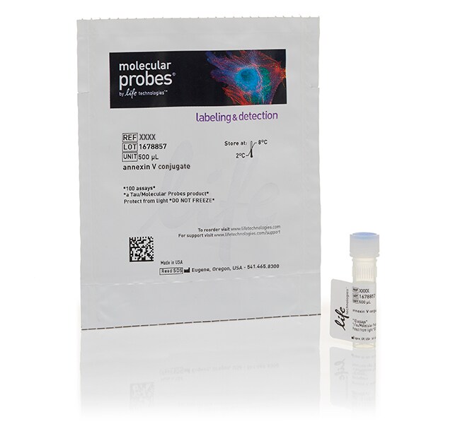
含 1 小瓶 Annexin V Alexa Fluor 647 偶联物。在冰箱(2°C 至 8°C)中避光储存。
常见问题解答 (FAQ)
我用胰蛋白酶消化贴壁细胞并用膜联蛋白V标记,现在甚至在对照的未处理细胞中,流式数据结果都显示出高比例的细胞凋亡,这是什么原因所致?
我能在成像实验中检测膜联蛋白V染色么?
流式细胞仪分析时,我应该在什么时候用膜联蛋白V染色粘连细胞?在胰蛋白酶消化前或后?
我能固定膜联蛋白V标记的细胞么?
我想用膜联蛋白V标记贴壁细胞,结果发现所有细胞都被标记了,该如何解决这一问题?
引用和文献 (30)
引用和文献
Abstract
The influenza A virus PB1-F2 protein targets the inner mitochondrial membrane via a predicted basic amphipathic helix that disrupts mitochondrial function.
Journal:J Virol
PubMed ID:12805420
'The 11th influenza A virus gene product is an 87-amino-acid protein provisionally named PB1-F2 (because it is encoded by an open reading frame overlapping the PB1 open reading frame). A significant fraction of PB1-F2 localizes to the inner mitochondrial membrane in influenza A virus-infected cells. PB1-F2 appears to enhance virus-induced
High-resolution mapping reveals topologically distinct cellular pools of phosphatidylserine.
Journal:J Cell Biol
PubMed ID:21788369
'Phosphatidylserine (PS) plays a central role in cell signaling and in the biosynthesis of other lipids. To date, however, the subcellular distribution and transmembrane topology of this crucial phospholipid remain ill-defined. We transfected cells with a GFP-tagged C2 domain of lactadherin to detect by light and electron microscopy PS exposed
Nuclear relocation of the nephrin and CD2AP-binding protein dendrin promotes apoptosis of podocytes.
Journal:Proc Natl Acad Sci U S A
PubMed ID:17537921
'Kidney podocytes and their slit diaphragms (SDs) form the final barrier to urinary protein loss. There is mounting evidence that SD proteins also participate in intracellular signaling pathways. The SD protein nephrin serves as a component of a signaling complex that directly links podocyte junctional integrity to actin cytoskeletal dynamics.
Transcriptional up-regulation of ULK1 by ATF4 contributes to cancer cell survival.
Journal:Biochem J
PubMed ID:23078367
'Hypoxia in the microenvironment of many solid tumours is an important determinant of malignant progression. The ISR (integrated stress response) protects cells from the ER (endoplasmic reticulum) stress caused by severe hypoxia. Likewise, autophagy is a mechanism by which cancer cells can evade hypoxic cell death. In the present paper
Phosphorylation of inositol 1,4,5-trisphosphate receptors by protein kinase B/Akt inhibits Ca2+ release and apoptosis.
Journal:Proc Natl Acad Sci U S A
PubMed ID:18250332
'Imbalance of signals that control cell survival and death results in pathologies, including cancer and neurodegeneration. Two pathways that are integral to setting the balance between cell survival and cell death are controlled by lipid-activated protein kinase B (PKB)/Akt and Ca(2+). PKB elicits its effects through the phosphorylation and inactivation