Search


引用和文献 (4)
Invitrogen™
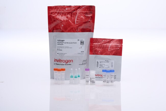
Alexa Fluor™ 647 微量蛋白标记试剂盒
微量蛋白标记试剂盒提供了一种便利的方法,可将荧光标记连接至少量抗体或蛋白 (20–100 µg)。试剂盒具有四种 Alexa Fluor™ 颜色(或生物素),提供三次标记和分离反应所需的全部试剂。微量蛋白标记试剂盒的重要特点:•标记蛋白通常可在了解更多信息
| 货号 | 数量 |
|---|---|
| A30009 | 1 个试剂盒 |
货号 A30009
价格(CNY)
10,713.00
Each
数量:
1 个试剂盒
价格(CNY)
10,713.00
Each
微量蛋白标记试剂盒提供了一种便利的方法,可将荧光标记连接至少量抗体或蛋白 (20–100 µg)。试剂盒具有四种 Alexa Fluor™ 颜色(或生物素),提供三次标记和分离反应所需的全部试剂。
微量蛋白标记试剂盒的重要特点:
•标记蛋白通常可在 2 小时内立即可用(手动操作时间 ∼30 分钟)
•针对分子量介于 12 和 150 kDa 之间的 20–100 µg 蛋白进行了优化
•使用便利的离心过滤柱进行纯化,得率介于 60% 和 90% 之间
•标记前必须去除样品中的稳定蛋白
稳定反应化学和卓越 Alexa Fluor™ 染料
在微量蛋白标记试剂盒中,反应性染料含有琥珀酰亚胺 (NHS) 酯部分,该部分与蛋白的伯胺反应形成稳定的染料-蛋白偶联物。与传统染料相比,Alexa Fluor™ 染料更明亮、光稳定性更好,且在 pH 值 4 至 10 之间的 pH 值耐受性更强。通常,当使用 Alexa Fluor™ 染料时,可达到更高程度的标记,而无需分子内淬灭。有关详细信息,请参见跨越可见光谱和红外光谱的 Alexa Fluor™ 染料—第 1.3 节。
了解更多关于蛋白和抗体标记的信息
我们可提供多种可供选择的 Molecular Probes™ 抗体和蛋白标记试剂盒,以适应您的起始材料和实验设置。参见 A 至 Z 的抗体标记或使用我们的标记化学选择工具进行其他选择。要了解更多关于不同试剂盒的信息,请阅读 Molecular Probes™ 手册中标记蛋白和核酸的试剂盒—第 1.2 节。
我们将为您定制抗体偶联物
如果您在我们的储存列表中找不到您想要的产品,我们将为您制备定制抗体偶联物。我们的定制偶联服务高效且保密, 而且我们绝对保证质量。我们获得了 ISO 9001:2000 认证。
仅供研究使用。不适用于动物或人类的治疗或诊断。
仅供科研使用。不可用于诊断程序。
规格
颜色远红外
检测方法荧光
激发/发射650/665
标签类型Alexa Fluor 染料
标记方法基于偶联
标记规模20 至 100 μg
产品线Alexa Fluor
产品类型蛋白标记试剂盒
数量1 个试剂盒
反应一部分琥珀酰亚胺 (NHS) 酯
运输条件室温
标记目标蛋白
标签或染料Alexa Fluor 647
Unit SizeEach
内容与储存
在冷藏冰箱(2°C 至 8°C)中避光储存。
常见问题解答 (FAQ)
我应该使用多大浓度的抗体进行偶联?
何为标记度(DOL)?
Can I use 50 μg of protein with Fluorescent Protein Labeling Kits?
What formulation of antibody should I use for conjugation for small animal in vivo imaging?
What is degree of labeling (DOL)?
引用和文献 (4)
引用和文献
Abstract
Ubiquitination screen using protein microarrays for comprehensive identification of Rsp5 substrates in yeast.
Journal:Molecular systems biology
PubMed ID:17551511
Ubiquitin-protein ligases (E3s) are responsible for target recognition and regulate stability, localization or function of their substrates. However, the substrates of most E3 enzymes remain unknown. Here, we describe the development of a novel proteomic in vitro ubiquitination screen using a protein microarray platform that can be utilized for the
Mouse marginal zone B cells harbor specificities similar to human broadly neutralizing HIV antibodies.
Journal:Proc Natl Acad Sci U S A
PubMed ID:23288906
'A series of potent, broadly neutralizing HIV antibodies have been isolated from B cells of HIV-infected individuals. VRC01 represents a subset of these antibodies that mediate neutralization with a restricted set of IGHV genes. The memory B cells expressing these antibodies were isolated years after infection; thus, the B-cell subpopulation
Microstructure, local viscoelasticity and cell culture suitability of 3D hybrid HA/collagen scaffolds.
Journal:PLoS One
PubMed ID:30566463
'As mechanical properties of cell culture substrates matter, methods for mechanical characterization of scaffolds on a relevant length scale are required. We used multiple particle tracking microrheology to close the gap between elasticity determined from bulk measurements and elastic properties sensed by cells. Structure and elasticity of macroporous, three-dimensional cryogel
Microtubule-Actomyosin Mechanical Cooperation during Contact Guidance Sensing.
Journal:Cell Rep
PubMed ID:30304674
Cancer cell migration through and away from tumors is driven in part by migration along aligned extracellular matrix, a process known as contact guidance (CG). To concurrently study the influence of architectural and mechanical regulators of CG sensing, we developed a set of CG platforms. Using flat and nanotextured substrates