Search

Invitrogen™
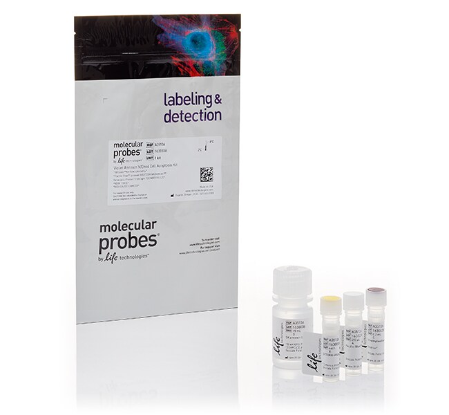
Pacific Blue™ 膜联蛋白 V/SYTOX™ AADvanced™ 细胞凋亡试剂盒,用于流式细胞分析
This product detects the externalization of phosphatidylserine in apoptotic cells using recombinant annexin V conjugated to violet-fluorescent Pacific Blue™ dye了解更多信息
| 货号 | 数量 |
|---|---|
| A35136 | 1 kit |
货号 A35136
价格(CNY)
4,499.00
飞享价
Ends: 31-Dec-2026
5,978.00共减 1,479.00 (25%)
Each
数量:
1 kit
价格(CNY)
4,499.00
飞享价
Ends: 31-Dec-2026
5,978.00共减 1,479.00 (25%)
Each
This product detects the externalization of phosphatidylserine in apoptotic cells using recombinant annexin V conjugated to violet-fluorescent Pacific Blue™ dye and dead cells using SYTOX™ AADvanced™ stain. After staining a cell population with Pacific Blue™, annexin V and SYTOX™ AADvanced™, apoptotic cells show violet fluorescence, dead cells show red fluorescence, and live cells show little or no fluorescence. These populations are easily distinguished by a flow cytometer with the 405 nm and 488 nm lines for excitation. There is very little spectral overlap between the two dyes, therefore very little compensation is needed. Each kit contains sufficient reagents for approximately 50 flow cytometry tests.
View a selection guide for all apoptosis assays for flow cytometry.
View a selection guide for all apoptosis assays for flow cytometry.
仅供科研使用。不可用于诊断程序。
规格
激发/发射Pacific Blue™:415⁄455,SYTOX™ AADvanced™:546⁄647
流式细胞仪激光线路405、488
适用于(应用)流式细胞分析
适用于(设备)流式细胞仪
反应次数50
产品类型细胞凋亡试剂盒
数量1 kit
运输条件湿冰
偶联物Pacific Blue™,SYTOX™ AADvanced™ 死细胞染色剂
产品规格管装
Unit SizeEach
内容与储存
含一小瓶膜联蛋白 V Pacific Blue™ 偶联物 (250 µL)、一小瓶 SYTOX™ AADvanced™ 死细胞染色、一瓶膜联蛋白结合缓冲液(5X 溶液,15 mL)和一小瓶 DMSO (100 µL)。
避光储存在冰箱 (2–8°C) 中。
避光储存在冰箱 (2–8°C) 中。