Search


引用和文献 (19)
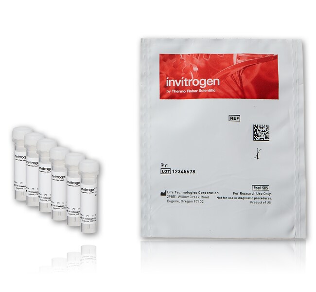
Invitrogen™
FluoSpheres™ 多尺寸规格试剂盒 #1,羧基修饰微球,红色荧光 (580/605),2% 固体,六种规格
Microspheres (also called latex beads or latex particles) are spherical particles in the colloidal size range that are formed from an amorphous polymer such as polystyrene.
| 货号 | 数量 |
|---|---|
| F8887 | 6 x 1 mL (1 mL/size) |
货号 F8887
价格(CNY)
13,863.00
Each
数量:
6 x 1 mL (1 mL/size)
价格(CNY)
13,863.00
Each
微球(又称乳胶微球或乳胶颗粒), 是胶体粒度范围内的球形颗粒, 由诸如聚苯乙烯等非晶态聚合物形成。我们的 Molecular Probes™ FluoSpheres™ 微珠采用高质量、超纯聚苯乙烯加工而成,标记有我们各种各样的特色染料,所形成的高强度荧光微珠即使在做荧光显微检测高强度激发照射时也很少、甚至不发生光漂白。该 FluoSpheres™ 多尺寸规格试剂盒µ针对以下规格提供 1 mL 溶液:0.02、0.1、0.2、0.5、1.0 和 2.0 m。
FluoSpheres™ 微球技术规格
• 标签(激发/发射波长):红色荧光 (580/605)
• 标称微珠直径:0.02、0.1、0.2、0.5、1.0、2.0 µm
• 偶联表面:羧酸盐
•固体:2%
不同 FluoSpheres™ 偶联表面的特性
• 羧基修饰 FluoSpheres™ 微珠的表面有高密度的侧链羧酸,这使得它们适于通过 EDAC 等水溶性碳化二亚胺试剂进行蛋白和其他含胺的生物分子的共价偶联。
•硫酸盐 FluoSpheres™ 微珠是相对疏水性颗粒,几乎不可逆地被动吸附所有蛋白,包括白蛋白、IgG、亲和素以及链霉亲和素。
•醛—硫酸盐 FluoSpheres™ 微珠表面添加醛基,旨在用于在非常温和的条件下与蛋白和其他胺发生反应。
•胺基改性 FluoSpheres™ 微珠利用水溶性的碳化二亚胺可与多种胺反应性分子偶联,包括琥珀酰亚胺酯及半抗原和药物的异硫氰酸盐或者蛋白的羧酸。
微球的关键应用
•仪器校准(流式细胞分析、显微镜、HTS、HCS)
•流量检测(微流体、血流量、水流量和空气流量)
•细胞生物学示踪剂(细胞分化和细胞示踪)
•免疫检测(凝集反应检测、ELISA、粒子捕获和对比度试剂)
选择 FluoSpheres™ 荧光微球
在我们提供的荧光微球完整产品系列中,您’将发现具有以下差异的微珠:
•10 种荧光颜色
• 10 种标称微珠直径:0.02 µm、0.04 µm、0.1 µm、0.2 µm、0.5 µm、1.0 µm、2.0 µm、4.0 µm、10.0 µm 和 15.0 µm
•蛋白偶联的四种表面改性:羧酸盐、硫酸盐、醛硫酸盐、胺
•另外与链霉素亲和素、NeutrAvidin、生物素、铕和铂预偶联的微球
未染色微球选择
我们还提供数百种 UltraClean™ 无表面活性剂的微球选项,用于研究和商业应用。
我们将为您定制微球产品’
我们将根据要求准备定制订单。例如,FluoSpheres™ 微珠可以低于我们常规的亮度,这在某些多色应用中非常适用。我们的定制结合服务是高效且保密的,我们保证工作的质量。我们获得了 ISO 9001:2000 认证。
仅供研究使用。不适用于动物或人类的治疗或诊断。
仅供科研使用。不可用于诊断程序。
规格
产品线FLUOSPHERES
数量6 x 1 mL (1 mL/size)
运输条件室温
表面改性羧基
颜色红色
直径(公制)2 μm、0.5 μm、0.1 μm、1 μm、0.02 μm、0.2 μm
材质聚苯乙烯
产品类型羧基修饰微球
Unit SizeEach
内容与储存
在冰箱(2°C 至 8°C)中避光储存。
常见问题解答 (FAQ)
洗涤和离心后,我的微球只留下了极少量的沉淀且溶液呈透明状。为什么会出现这种现象?
我的微球已保存超过一年,我想知道它们是否仍可正常使用,有什么好办法来核实它们的功能是否完好?
我不慎冷冻了自己的微球,还可继续使用么?
在我的实验体系中,蛋白包被的微球出现非特异性结合。有什么产品可以帮助减少这些非特异性作用吗?
我应该使用哪种方法来分散聚集体?
引用和文献 (19)
引用和文献
Abstract
Tuftsin binds neuropilin-1 through a sequence similar to that encoded by exon 8 of vascular endothelial growth factor.
Journal:The Journal of biological chemistry
PubMed ID:16371354
Syntaxin 7 is localized to late endosome compartments, associates with Vamp 8, and Is required for late endosome-lysosome fusion.
Journal:Mol Biol Cell
PubMed ID:10982406
'Protein traffic from the cell surface or the trans-Golgi network reaches the lysosome via a series of endosomal compartments. One of the last steps in the endocytic pathway is the fusion of late endosomes with lysosomes. This process has been reconstituted in vitro and has been shown to require NSF,
Fractal nature of regional ventilation distribution.
Journal:J Appl Physiol
PubMed ID:10797111
'High-resolution measurements of pulmonary perfusion reveal substantial spatial heterogeneity that is fractally distributed. This observation led to the hypothesis that the vascular tree is the principal determinant of regional blood flow. Recent studies using aerosol deposition show similar ventilation heterogeneity that is closely correlated with perfusion. We hypothesize that ventilation
Mapping mechanical strain of an endogenous cytoskeletal network in living endothelial cells.
Journal:Biophys J
PubMed ID:12668477
'A central aspect of cellular mechanochemical signaling is a change of cytoskeletal tension upon the imposition of exogenous forces. Here we report measurements of the spatiotemporal distribution of mechanical strain in the intermediate filament cytoskeleton of endothelial cells computed from the relative displacement of endogenous green fluorescent protein (GFP)-vimentin before
Viewing dynamic assembly of molecular complexes by multi-wavelength single-molecule fluorescence.
Journal:Biophys J
PubMed ID:16698779
'Complexes of macromolecules that transiently self-assemble, perform a particular function, and then dissociate are a recurring theme in biology. Such systems often have a large number of possible assembly/disassembly intermediates and complex, highly branched reaction pathways. Measuring the single-step kinetic parameters in these reactions would help to identify the functionally