Search


引用和文献 (10)
Invitrogen™
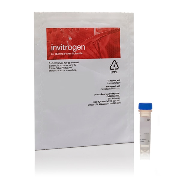
pHrodo™ Red 和 Green 表皮生长因子 (EGF) 和 pHrodo™ Red 转铁蛋白偶联物
利用 pHrodo Red 和 pHrodo Green 表皮生长因子 (EGF) 和 pHrodo Red 转铁蛋白偶联物,可在 HCS、流式细胞分析、基于微孔板的荧光测定和 HTS 等多种应用中用于检测细胞中的内化 EGF 和转铁蛋白。
Have Questions?
更改视图

| 货号 | 产品类型 |
|---|---|
| P35376 | 细胞标记溶液 |
| P35374 | 细胞标记溶液 |
货号 P35376
价格(CNY)
6,431.00
Each
产品类型:
细胞标记溶液
价格(CNY)
6,431.00
Each
利用具有高活性的 pHrodo Red 和 pHrodo Green 表皮生长因子 (EGF) 偶联物和 pHrodo Red 转铁蛋白偶联物检测内化的 EGF 或转铁蛋白。这些偶联物包括与高活性 EGF 或转铁蛋白分子直接关联的 pHrodo 染料,可在 HCS、流式细胞分析、微孔板荧光测定和 HTS 等多种应用中用于检测内化的 EGF 和转铁蛋白分子。pHrodo Red 和 pHrodo Green 染料在暴露于酸性环境(如内体和溶酶体中)时呈强荧光。
pHrodo Red 和 pHrodo Green 表皮生长因子 (EGF) 偶联物由 pHrodo Red 或 pHrodo Green 染料组成,可直接偶联高活性的人类重组 EGF 分子并用于检测细胞中的内化 EGF 受体。荧光标记的 EGF 使科学家能够考察受体-膜之间的相互作用,研究受体分布,计算 EGF 与受体相互作用的速率常数等。随 pH 值变化而变化的荧光标记进一步提高了检测水平。
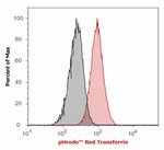
pHrodo Red 转铁蛋白偶联物由 pHrodo Red 染料组成,可直接偶联高活性转铁蛋白分子,用于检测细胞中的内化转铁蛋白。转铁蛋白是一种单体血清糖蛋白(∼80,000 道尔顿),可结合脊椎动物细胞表面的特异性受体,并通过受体介导的内吞作用,递送多达两个 Fe3+ 原子。携带铁的转铁蛋白进入核内体后,酸性环境即可促使铁离子从转铁蛋白–受体复合物中解离。铁释放后,脱铁转铁蛋白被回收至细胞膜中,其从受体中释放出来以结合更多的铁。
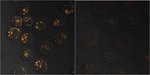
高度光稳定的 pHrodo Red 和 pHrodo Green 染料在中性 pH 条件下呈弱荧光,但是当通过 EGF 或转铁蛋白特异性内化时,在酸性内体中呈强荧光。因此,其可测定内化的 EGF 或转铁蛋白以及动态监测活细胞中的 EGF 或转铁蛋白内化过程。
pHrodo Red EGF 偶联物和 pHrodo Red 转铁蛋白偶联物的激发/发射波长为 560/585 nm,可与多种蓝色、红色和远红色染料报告基因(如 Mitosox Red、CellEvent Caspase 3/7 Red、NucBlue、RFP 和 Mitotracker Deep Red 等)配合使用进行多通路检测。pHrodo Green EGF 偶联物的激发/发射波长为 509/533 nm,可与多种蓝色、红色和远红色染料报告基因(如 Mitosox Red、CellEvent Caspase 3/7 Red、NucBlue、RFP 和 Mitotracker Deep Red 等)配合使用进行多通路检测。
pHrodo Red 与 pHrodo Green EGF 偶联物和 pHrodo Red 转铁蛋白偶联物与多种应用兼容,包括传统荧光显微术、高内涵筛选 (HCS)、流式细胞分析和基于微孔板的荧光测定或高通量筛选 (HTS) 等。这些偶联物还与各种宽视野或共聚焦荧光显微镜、流式细胞仪、荧光酶标仪和高内涵分析仪兼容。
pHrodo Red 转铁蛋白偶联物由 pHrodo Red 染料组成,可直接偶联高活性转铁蛋白分子,用于检测细胞中的内化转铁蛋白。转铁蛋白是一种单体血清糖蛋白(∼80,000 道尔顿),可结合脊椎动物细胞表面的特异性受体,并通过受体介导的内吞作用,递送多达两个 Fe3+ 原子。携带铁的转铁蛋白进入核内体后,酸性环境即可促使铁离子从转铁蛋白–受体复合物中解离。铁释放后,脱铁转铁蛋白被回收至细胞膜中,其从受体中释放出来以结合更多的铁。
高度光稳定的 pHrodo Red 和 pHrodo Green 染料在中性 pH 条件下呈弱荧光,但是当通过 EGF 或转铁蛋白特异性内化时,在酸性内体中呈强荧光。因此,其可测定内化的 EGF 或转铁蛋白以及动态监测活细胞中的 EGF 或转铁蛋白内化过程。
pHrodo Red EGF 偶联物和 pHrodo Red 转铁蛋白偶联物的激发/发射波长为 560/585 nm,可与多种蓝色、红色和远红色染料报告基因(如 Mitosox Red、CellEvent Caspase 3/7 Red、NucBlue、RFP 和 Mitotracker Deep Red 等)配合使用进行多通路检测。pHrodo Green EGF 偶联物的激发/发射波长为 509/533 nm,可与多种蓝色、红色和远红色染料报告基因(如 Mitosox Red、CellEvent Caspase 3/7 Red、NucBlue、RFP 和 Mitotracker Deep Red 等)配合使用进行多通路检测。
pHrodo Red 与 pHrodo Green EGF 偶联物和 pHrodo Red 转铁蛋白偶联物与多种应用兼容,包括传统荧光显微术、高内涵筛选 (HCS)、流式细胞分析和基于微孔板的荧光测定或高通量筛选 (HTS) 等。这些偶联物还与各种宽视野或共聚焦荧光显微镜、流式细胞仪、荧光酶标仪和高内涵分析仪兼容。
仅供科研使用。不可用于诊断程序。
规格
描述pHrodo™ Red 转铁蛋白偶联物
检测方法荧光
染料类型pHrodo Red
激发/发射560/585
形式冻干粉
数量1 mg
子细胞定位核内体
颜色橙色
适用于(应用)细胞分析
适用于(设备)荧光显微镜, 流式细胞仪, Qubit™ 2.0定量仪, 高内涵分析仪
产品线pHrodo
产品类型细胞标记溶液
Unit SizeEach
内容与储存
在 –5°C 至 –30°C 下干燥环境中避光储存。
引用和文献 (10)
引用和文献
Abstract
Identification of epithelial ovarian tumor-specific aptamers.
Journal:
PubMed ID:25894736
'Ovarian cancer is often diagnosed in late stages with few treatment options and poor long-term prognosis. New clinical tools for early detection of ovarian malignancies will significantly help reduce mortality and improve current long-term survival rates. The objective of this work was to identify ovarian tumor-specific single-stranded DNA aptamers that
Scavenger receptor-recognized and enzyme-responsive nanoprobe for fluorescent labeling of lysosomes in live cells.
Journal:
PubMed ID:24929616
Lysosomal imaging represents a potent tool for investigating the organization of related cellular events and their modulation via diagnostic and therapeutic approaches. However, specific labeling of the lysosome in live cells is a significant challenge. Taking advantage of the inherent lysosomal entry of nanoparticles and unique digestive inclusions in the
Systematic mapping of genetic interactions for de novo fatty acid synthesis identifies C12orf49 as a regulator of lipid metabolism.
Journal:Nat Metab
PubMed ID:32694731
Synthesis and Biological Characterization of a New Norbormide Derived Bodipy FL-Conjugated Fluorescent Probe for Cell Imaging.
Journal:Front Pharmacol
PubMed ID:30319407
LY6E mediates an evolutionarily conserved enhancement of virus infection by targeting a late entry step.
Journal:Nat Commun
PubMed ID:30190477