Search


引用和文献 (13)
Invitrogen™
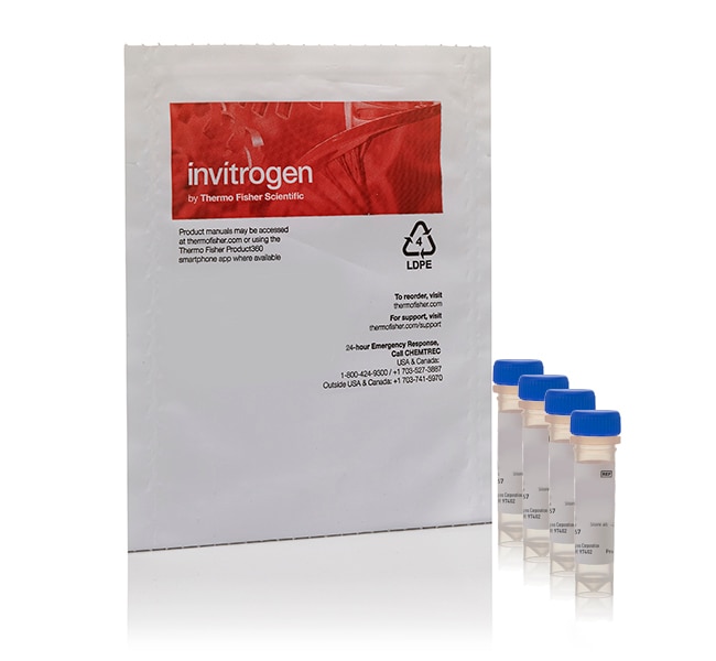
PS-Speck™显微镜点源试剂盒(蓝色、绿色、橙色 & 深红色荧光微珠)
PS-Speck™显微镜点源试剂盒含有四种不同颜色的荧光微球;每个微球的直径为 0.175 +/- 0.005 μm。这种格外小的直径变异系数使 PS-Speck™ 微球适用于校准仪器光学器件的均匀、亚分辨率荧光源,特别是在三维成像应用中。该试剂盒的四种即用型了解更多信息
| 货号 | 数量 |
|---|---|
| P7220 | 1 kit |
货号 P7220
价格(CNY)
3,839.00
Each
数量:
1 kit
价格(CNY)
3,839.00
Each
PS-Speck™显微镜点源试剂盒含有四种不同颜色的荧光微球;每个微球的直径为 0.175 +/- 0.005 μm。这种格外小的直径变异系数使 PS-Speck™ 微球适用于校准仪器光学器件的均匀、亚分辨率荧光源,特别是在三维成像应用中。该试剂盒的四种即用型 1 mL 悬浮液含有明亮的单分散微球,其激发/发射波长为 360/440 nm(蓝色)、505/515 nm(绿色)、540/560 nm(橙色)和 633/660 nm(深红色)。PS-Speck™ 试剂盒还包含封片方案,便于用户使用,并提供足够的封片剂,可各制备 100 个载玻片。
查看我们完整的显微镜校准试剂系列›
查看我们完整的显微镜校准试剂系列›
仅供科研使用。不可用于诊断程序。
规格
校准类型光学校准,荧光显微镜校准
适用于(应用)内部或外部荧光显微镜检查标准品
产品规格悬浮微珠
数量1 kit
运输条件室温
颜色橙色、深红色、蓝色、绿色
产品线PS-Speck
类型显微镜点源试剂盒
Unit SizeEach
内容与储存
请避光冷藏 (2–8°C) 储存。
常见问题解答 (FAQ)
What is the refractive index of the mounting medium in PS-Speck Microscope Point Source Kit (Cat. No. P7220)?
引用和文献 (13)
引用和文献
Abstract
Correcting confocal acquisition to optimize imaging of fluorescence resonance energy transfer by sensitized emission.
Journal:Biophys J
PubMed ID:15041688
Imaging of fluorescence resonance energy transfer (FRET) between suitable fluorophores is increasingly being used to study cellular processes with high spatiotemporal resolution. The genetically encoded Cyan (CFP) and Yellow (YFP) variants of Green Fluorescent Protein have become the most popular donor and acceptor pair in cell biology. FRET between these
In vivo mammalian brain imaging using one- and two-photon fluorescence microendoscopy.
Journal:J Neurophysiol
PubMed ID:15128753
'One of the major limitations in the current set of techniques available to neuroscientists is a dearth of methods for imaging individual cells deep within the brains of live animals. To overcome this limitation, we developed two forms of minimally invasive fluorescence microendoscopy and tested their abilities to image cells
Interaction of Maf transcription factors with Pax-6 results in synergistic activation of the glucagon promoter.
Journal:J Biol Chem
PubMed ID:11457839
'In the endocrine pancreas, alpha-cell-specific expression of the glucagon gene is mediated by DNA-binding proteins that interact with the G1 proximal promoter element. Among these proteins, the paired domain transcription factor Pax-6 has been shown to bind to G1 and to transactivate glucagon gene expression. Close to the Pax-6-binding site,
Quality assessment of confocal microscopy slide based systems: performance.
Journal:Cytometry A
PubMed ID:16807897
'BACKGROUND: All fluorescence slide-based cytometry detections systems basically include the following components: (1) an excitation light source, (2) intermediate optics, and (3) a detection device consisting of a CCD camera or a PMT. The optical principles employed is slide-based systems are similar to those of confocal microscopes (CLSM). METHODS: The
Spatial organization of bacteriorhodopsin in model membranes. Light-induced mobility changes.
Journal:J Biol Chem
PubMed ID:12167614
'Bacteriorhodopsin is a proton-transporting membrane protein in Halophilic archaea, and it is considered a prototype of membrane transporters and a model for G-protein-coupled receptors. Oligomerization of the protein has been reported, but it is unknown whether this feature is correlated with, for instance, light activation. Here, we have addressed this