Search


引用和文献 (10)
Invitrogen™
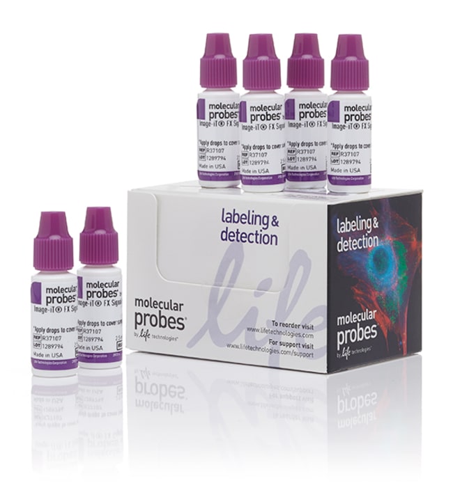
Image-iT™ FX 信号增强剂 ReadyProbes™ 试剂
Image-iT™ FX Signal Enhancer ReadyProbes™ Reagent dramatically reduces background fluorescence resulting from non-specific staining during secondary detection using conjugates of streptavidin or secondary antibodies.
| 货号 | 数量 |
|---|---|
| R37107 | 6瓶 |
货号 R37107
价格(CNY)
2,864.00
Online exclusive
Ends: 31-Dec-2026
3,977.00共减 1,113.00 (28%)
Each
数量:
6瓶
价格(CNY)
2,864.00
Online exclusive
Ends: 31-Dec-2026
3,977.00共减 1,113.00 (28%)
Each
Image-iT™ FX Signal Enhancer ReadyProbes™ Reagent dramatically reduces background fluorescence resulting from non-specific staining during secondary detection using conjugates of streptavidin or secondary antibodies. This reagent is applied directly to slides or coverslips containing fixed and permeabilized cell or tissue samples prior to staining with fluorescent probes.
- Easy protocol – apply ready-made solution using a dropper bottle
- Compatible with the other steps in your immunostaining protocol
- Effective with most fluorescent labels
- Stable at room temperature – keep handy at your scope or cell culture area
Image-IT™ FX 信号增强剂 ReadyProbes™ 试剂可大幅降低使用链霉亲和素或二抗偶联物进行二次检测期间因非特异性染色而产生的背景荧光。该试剂可在用荧光探针染色之前直接应用于包被固定和透化的细胞或组织样品的载玻片或盖玻片。
•操作简单—使用滴管添加现成的溶液
• 与免疫染色方案中的其他步骤兼容
• 对大多数荧光标记有效
• 室温下稳定—可放置在视野范围内或细胞培养区域,方便取用
查看其他 ReadyProbes™ 细胞染色试剂
查找更多固定细胞成像工具
细胞成像应用
Image-iT™ FX 信号增强剂是一种可有效阻断背景染色的产品,这种背景染色是由多种荧光染料与细胞和组织成分之间的非特异性相互作用导致的。在染色前对固定、透化的细胞采用 Image-iT™ FX 信号增强剂时,可在很大程度上消除因使用链霉亲和素荧光偶联物和抗体偶联物而产生的背景染色。
使用建议
•在进行免疫染色之前,将 Image-iT™ FX 信号增强剂直接应用于包被细胞或组织样品的载玻片或盖玻片。
•固定/透化步骤后,只需清洗、添加足够覆盖样品的液滴,孵育 30 分钟然后进行正常的染色操作。
•根据需要,在使用 Image-iT™ 信号增强剂 ReadyProbes™ 试剂封闭后可进行其他封闭步骤
•操作简单—使用滴管添加现成的溶液
• 与免疫染色方案中的其他步骤兼容
• 对大多数荧光标记有效
• 室温下稳定—可放置在视野范围内或细胞培养区域,方便取用
查看其他 ReadyProbes™ 细胞染色试剂
查找更多固定细胞成像工具
细胞成像应用
Image-iT™ FX 信号增强剂是一种可有效阻断背景染色的产品,这种背景染色是由多种荧光染料与细胞和组织成分之间的非特异性相互作用导致的。在染色前对固定、透化的细胞采用 Image-iT™ FX 信号增强剂时,可在很大程度上消除因使用链霉亲和素荧光偶联物和抗体偶联物而产生的背景染色。
使用建议
•在进行免疫染色之前,将 Image-iT™ FX 信号增强剂直接应用于包被细胞或组织样品的载玻片或盖玻片。
•固定/透化步骤后,只需清洗、添加足够覆盖样品的液滴,孵育 30 分钟然后进行正常的染色操作。
•根据需要,在使用 Image-iT™ 信号增强剂 ReadyProbes™ 试剂封闭后可进行其他封闭步骤
仅供科研使用。不可用于诊断程序。
规格
产品规格Bottle(s)
产品线Image-iT、ReadyProbes
数量6瓶
产品类型FX 信号增强试剂
溶液类型信号增强剂
Unit SizeEach
内容与储存
2.5 mL 滴瓶
在 ≤ 25°C 下储存。
在 ≤ 25°C 下储存。
常见问题解答 (FAQ)
当我分析click标记样品时,观测到较高的本底干扰,这是什么原因所致?如何减少本底干扰?
我用了一种神经元特异性抗体标记我的神经元,怎样才能减少非特异性抗体结合?
我在对照细胞的细胞核和线粒体中看到非特异性结合,但是蛋白质结合不足以将其终止。我应该怎么做?
I used Image-iT FX Signal Enhancer solution to get rid of nonspecific nuclear labeling with Alexa Fluor 568 secondary antibody, but I also saw a significant reduction in my specific mitochondrial antibody labeling. Why is this and what can I do?
Can I use the Image-iT FX Signal Enhancer instead of my normal blocking solution (BSA or serum)?
引用和文献 (10)
引用和文献
Abstract
Functional scaffold-free 3-D cardiac microtissues: a novel model for the investigation of heart cells.
Journal:Am J Physiol Heart Circ Physiol
PubMed ID:22427522
To bridge the gap between two-dimensional cell culture and tissue, various three-dimensional (3-D) cell culture approaches have been developed for the investigation of cardiac myocytes (CMs) and cardiac fibroblasts (CFs). However, several limitations still exist. This study was designed to develop a cardiac 3-D culture model with a scaffold-free technology
Generation of an induced pluripotent stem cell line (TRNDi008-A) from a Hunter syndrome patient carrying a hemizygous 208insC mutation in the IDS gene.
Journal:Stem Cell Res
PubMed ID:31071499
'Mucopolysaccharidosis Type II (MPS II), also known as Hunter syndrome, is a rare X-linked genetic disease caused by mutations in the IDS gene encoding iduronate 2-sulfatase (I2S). This is a multisystem disorder with significant variation in symptoms. Here, we document a human induced pluripotent stem cell (iPSC) line generated from
Three-dimensional residual channel attention networks denoise and sharpen fluorescence microscopy image volumes.
Journal:Nat Methods
PubMed ID:34059829
The aberrant upregulation of exon 10-inclusive SREK1 through SRSF10 acts as an oncogenic driver in human hepatocellular carcinoma.
Journal:Nat Commun
PubMed ID:35296659
HIV-infected macrophages resist efficient NK cell-mediated killing while preserving inflammatory cytokine responses.
Journal:Cell Host Microbe
PubMed ID:33571449