How do SYTO dyes bind to DNA?
The binding mode of SYTO nucleic acid stains is unknown. However, the behavior of these and related nucleic acid dyes suggests the following binding properties:
1.They appear to contact the solvent (suggested by sensitivity to salt, divalent cations, and in particular, SDS) and thus are likely to have contacts in the grooves.
2.All SYTO dyes appear to show some base selectivity and are thus likely to have minor groove contacts.
3.They can be removed from nucleic acid via ethanol precipitation; this characteristic is not shared by ethidium bromide and other intercalators. Likewise, the dyes are not removed from nucleic acid via butanol or chloroform extraction. These extraction methods do remove ethidium bromide from nucleic acid.
4. SYTO binding is not affected by nonionic detergents.
5. SYTO dyes are not quenched by BrdU, so they do not bind nucleic acids in precisely the same way as Hoechst 33342 and DAPI ((4′,6-diamidino-2-phenylindole).
SYBR Green I has shown little mutagenicity on frameshift indicator strains, indicating that it isn't likely to strongly intercalate.
Find additional tips, troubleshooting help, and resources within our Cell Analysis Support Center.
I am using SYTOX AAdvanced as a dead cell stain, but all of my cells are labeling even though I am certain that they are supposed to be alive. These are adherent cells that I have trypsinized. Why am I getting false-dead signals?
SYTOX AAdvanced labels only dead cells because it is a cell impermeant dye. The dye can only enter cells that have a compromised plasma membrane. Trypsinization may cause temporary disruption of the plasma membrane, sufficient to allow staining with a cell impermeant dye. You can reduce the “false-dead” problem by either reducing the amount of trypsin and/or reduce the incubation time for trypsinization or use a gentler dissociation reagent such as TrypLE Express, TrypLESelect reagents, or Versene. After trypsinization, wash well, and if possible, allow a recovery time in normal culture media before staining with any of the SYTOX dyes.
Find additional tips, troubleshooting help, and resources within our Cell Analysis Support Center.
What kinds of cell health and viability assays can be performed by flow cytometry?
The following cell health and viability assays can be performed by flow cytometry :
-Apoptosis Assays:
Membrane Asymmetry: Annexin V is a member of a family of structurally related proteins that bind phospholipids in the presence of Ca2+. Annexin V binds several phospholipids, but shows highest affinity for phosphatidylserine.
Phosphatidylserine is normally found in the inner leaflet of the cell membrane; however, in the early stages of apoptosis, phosphatidylserine is observed to translocate to the outer leaflet. This translocation makes phosphatidylserine available for annexin V binding in the presence of Ca2+ containing incubation buffer. Cells undergoing apoptosis will stain with annexin V, while normal cells will not. annexin V is available conjugated with a wide range of fluorophores.
Mitochondrial Health: A distinctive feature of the early stages of apoptosis is the disruption of the mitochondria, including changes in membrane and redox potential. We exclusively offer a number of fluorescent probes for analyzing mitochondrial activity in live cells by flow cytometry, with minimal disruption of cellular function.
The MitoProbe family of mitochondrial stains (MitoProbe DiOC2(3) Assay Kit, Cat. No. M34150, MitoProbe JC-1 Assay Kit, Cat. No. M34152, and MitoProbe DiIC1(5) Assay Kit, Cat. No. M34151) provides quick, easy, and reliable flow cytometric detection of the loss of mitochondrial membrane potential that occurs during apoptosis.
Caspase Activity: The CellEvent Caspase-3/7 Green Flow Cytometry Assay Kit (Cat. No. C10427) enables flow cytometric detection of activated caspase-3 and caspase-7 in apoptotic cells. The kit includes the novel fluorogenic substrate CellEvent Caspase-3/7 Green Detection Reagent which targets the recognition sequence for activated caspase-3 and caspase-7, as well as SYTOX AADvanced Dead Cell Stain.
DNA Fragmentation: The later stages of apoptosis are characterized by changes in nuclear morphology, including DNA fragmentation, chromatin condensation, degradation of nuclear envelope, nuclear blebbing, and DNA strand breaks. DNA fragmentation that occurs during apoptosis produces DNA strand breaks, and can be analyzed using TUNEL (terminal deoxynucleotidyl transferase dUTP nick end labeling) assays. The APO-BrdU TUNEL assay (Cat. No. A23210) is a two-color assay for labeling DNA breaks and total cellular DNA to detect apoptotic cells by imaging or flow cytometry.
Nuclear Chromatin Condensation: The later stages of apoptosis are characterized by changes in nuclear morphology, including DNA fragmentation, chromatin condensation, degradation of nuclear envelope, nuclear blebbing, and DNA strand breaks. Cells undergoing apoptosis display an increase in nuclear chromatin condensation. As the chromatin condenses, cell-permeable nucleic acid stains becomes hyperfluorescent, thus enabling the identification of apoptotic cells when combined with a traditional dead-cell stain. The Vybrant Apoptosis Assay Kit #5, Hoechst 33342/Propidium Iodide (Cat. No. V13244) provides a rapid and convenient assay for apoptosis based on fluorescence detection of the compacted state of the chromatin in apoptotic cells. The Chromatin Condensation & Membrane Permeability Dead Cell Apoptosis Kit with Hoechst 33342, YO-PRO-1, and PI dyes, for flow cytometry (Cat. No. V23201) detects apoptotic cells with changes in nuclear chromatin condensation and plasma membrane permeability.
-Cell Cycle Analysis:
Live cell assays: The Vybrant DyeCycle family of dyes offers robust fluorescent dyes for live-cell cycle analysis with limited cytotoxicity using 405 nm (Cat. No. V35003), 488 nm (Cat. No. V35004), 532 nm (Cat. No. V35005), or 633 nm (Cat. Nos. V10309 and V10273) excitation. The dyes have low cytotoxicity, allowing stained cells to be sorted and otherwise cultured or assessed with functional assays after staining.
Fixed cell assays: Analyzing cell cycle using FxCycle Violet Stain (Cat. No. F10347), SYTOX AADvanced Dead Cell Stain Kit (Cat. No. S10349) or FxCycle Far Red Stain (Cat. No. F10348) allows for multiple color options for simplified fixed cell cycle analysis.
-Cell Proliferation:
Dye dilution assays for cell proliferation: Dye dilution assays for cell proliferation rely on cell membrane–permeant fluorescent molecules. Upon entry into the cell, the dye will covalently bind to amine groups on proteins, resulting in long-term dye retention within the cell. Through subsequent cell divisions, each daughter cell receives approximately half the fluorescence of the parent. Analysis of the fluorescence intensities of cell populations by flow cytometry enables determination of the number of generations through which a cell or population has progressed since the label was applied. CellTrace fluorescent stains can be used without affecting morphology or physiology to trace generations in vivo or in vitro. There is no known effect on proliferative ability or biology of cells and they are well retained in cells for several days post-stain. Available kits for flow cytometry include CellTrace CFSE Cell Proliferation Kit (Cat. No. C34554), CellTrace Violet Cell Proliferation Kit (Cat. No. C34557), and CellTrace Far Red Cell Proliferation Kit (Cat. No. C34564).
DNA Synthesis Assays: Measuring the synthesis of new DNA is a precise way to assay cell proliferation in individual cells or in cell populations. DNA synthesis–based cell proliferation assays measure the rate of new DNA synthesis based on incorporation of modified nucleosides. The Click-iT Plus EdU cell proliferation assay utilizes the power of click chemistry and the modified nucleoside EdU to provide a superior alternative to BrdU staining for detecting and quantitating newly synthesized DNA. The Click-iT Plus EdU cell proliferation assay is available with Pacific Blue (Cat. No. C10636), Alexa Fluor 488 (Cat. Nos. C10632 and C10633), and Alexa Fluor 647 (Cat. Nos. C10634 and C10635).
-Viability Assays:
Dead cells often give false positive results, as they tend to bind non-specifically to many reagents. Therefore, removing dead cells from your flow cytometry data is a critical step to help ensure accurate results and analysis.
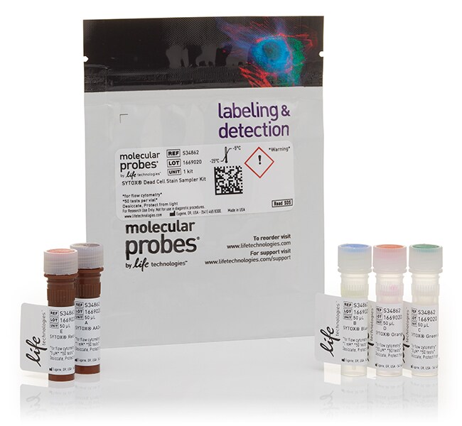
Non-fixable Membrane Permeability Stains: SYTOX Dead Cell Stains (Cat. Nos. S34857, S34860, S34861, S34859, and S34862) do not cross intact cell membranes, and they exhibit increased fluorescence upon dsDNA binding, making them some of our most brilliant dead cell stains. Cell-impermeant classic DNA-binding dyes include propidium iodide (Cat. No. P21493) and 7-AAD (Cat. No. A1310). Both of these dyes have been used extensively for viability assays in flow cytometry. CellTrace Calcein AM dyes can be passively loaded into adherent and nonadherent cells. These cell-permeant esterase substrates serve as viability probes that measure both enzymatic activity, which is required to activate their fluorescence, and cell membrane integrity, which is required for intracellular retention of their fluorescent products. Available with blue (Cat. No. C34853), violet (Cat. No. C34858), and green (Cat. No. C34852) fluorescence, these dyes are ideal for short-term staining of live cells and can be used in multiplexed flow cytometry experiments.
Fixable Viability Stains: The LIVE/DEAD Fixable Dead Cell Stains are fixable viability dyes that help to ensure accurate assessment of cell viability in samples after fixation and/or permeabilization. LIVE/DEAD Fixable Dead Cell Stain Kits are based on the reaction of a fluorescent reactive dye with cellular proteins (amines). These dyes cannot penetrate live-cell membranes, so only cell-surface proteins are available to react with the dye, resulting in dim staining. The reactive dye can permeate the damaged membranes of dead cells and stain both the interior and exterior amines, resulting in more intense staining. LIVE/DEAD Fixable Dead Cell Stain Kits are available in eight single-channel colors available for UV, 405, 488, 532, 561, or 633 nm lasers in three packaging sizes to match your experiment.
Find additional tips, troubleshooting help, and resources within our Cell Analysis Support Center.