Search


引用和文献 (16)
Invitrogen™
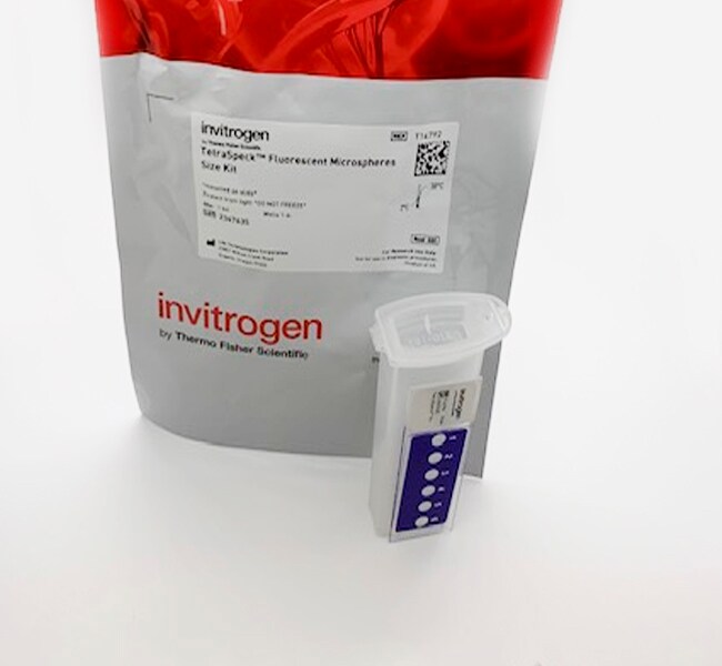
TetraSpeck™ 多尺寸荧光微球套件(固定在载玻片上)
TetraSpeck 多尺寸荧光微球套件包含一张显微镜载玻片,上有6个观察区。其中5个观察区各含有一种尺寸的微球 – 0.1、0.2、0.5、1.0或4.0 μm。其余区域含有5种尺寸的微球了解更多信息
| 货号 | 数量 |
|---|---|
| T14792 | 1 Kit |
货号 T14792
价格(CNY)
4,637.00
飞享价
Ends: 31-Dec-2026
6,266.00共减 1,629.00 (26%)
1 kit
数量:
1 Kit
价格(CNY)
4,637.00
飞享价
Ends: 31-Dec-2026
6,266.00共减 1,629.00 (26%)
1 kit
TetraSpeck 多尺寸荧光微球套件包含一张显微镜载玻片,上有6个观察区。其中5个观察区各含有一种尺寸的微球 – 0.1、0.2、0.5、1.0或4.0 μm。其余区域含有5种尺寸的微球。每个微球都使用四种不同的荧光染料 [365⁄430 nm(蓝色)、505⁄515 nm(绿色)、560⁄580 nm(橙色)和660⁄680 nm(深红色)] 染色,它们的激发峰和发射峰完全分开。Invitrogen’s TetraSpeck™荧光微球有助于校准宽场、TIRF 和共聚焦荧光显微镜,特别是用于多色应用,如共定位分析和对反卷积至关重要的点扩散函数的推导。
查看我们的全套显微镜校准试剂 ›
查看我们的全套显微镜校准试剂 ›
仅供科研使用。不可用于诊断程序。
规格
校准类型共聚焦显微镜校准、荧光显微镜校准
产品规格固定在载玻片上
产品线TetraSpeck
数量1 Kit
运输条件室温
颜色橙色、深红色、蓝色、绿色, Dark Red, Blue, Green
直径(公制)0.5 μm、0.2 μm、0.1 μm、4 μm、1 μm
产品类型荧光微球尺寸试剂盒
Unit Size1 kit
内容与储存
室温避光储存。
常见问题解答 (FAQ)
What are the excitation/emission peaks for TetraSpeck Microspheres?
The TetraSpeck Microspheres (Cat. Nos. T7279, T7280, T7281, T7283, T7284, T14792) are stained throughout with four different fluorescent dyes, yielding beads that each display four well-separated excitation/emission peaks at 360/430 nm (blue), 505/515 nm (green), 560/580 nm (orange) and 660/680 nm (dark red).
TetraSpeck Blue Dye Spectra
Fluorescence excitation and emission spectra of bead encapsulated TetraSpeck blue dye.

TetraSpeck Orange Dye Spectra

TetraSpeck Green Dye Spectra

TetraSpeck Dark Red Dye Spectra

Find additional tips, troubleshooting help, and resources within our Cell Analysis Support Center.
引用和文献 (16)
引用和文献
Abstract
The cohesion protein ORD is required for homologue bias during meiotic recombination.
Journal:J Cell Biol
PubMed ID:15007062
'During meiosis, sister chromatid cohesion is required for normal levels of homologous recombination, although how cohesion regulates exchange is not understood. Null mutations in orientation disruptor (ord) ablate arm and centromeric cohesion during Drosophila meiosis and severely reduce homologous crossovers in mutant oocytes. We show that ORD protein localizes along
Exocytosis of IgG as mediated by the receptor, FcRn: an analysis at the single-molecule level.
Journal:Proc Natl Acad Sci U S A
PubMed ID:15258288
'IgG transport within and across cells is essential for effective humoral immunity. Through a combination of biochemical and in vivo analyses, the MHC class I-related neonatal Fc receptor (FcRn) is known to play a central role in delivering IgGs within and across cells. However, little is known about the molecular
Colocalization of fluorescent markers in confocal microscope images of plant cells.
Journal:Nat Protoc
PubMed ID:18388944
'This protocol describes the steps needed to perform quantitative statistical colocalization on two-color confocal images, specifically of plant cells. The procedure includes a calibration test to check the chromatic alignment of the confocal microscope. A software tool is provided to calculate the Pearson and Spearman correlation coefficients (''Pearson-Spearman correlation colocalization''
Mast cell degranulation requires N-ethylmaleimide-sensitive factor-mediated SNARE disassembly.
Journal:J Immunol
PubMed ID:14607937
'Mast cells possess specialized granules that, upon stimulation of surface FcR with IgE, fuse with the plasma membrane, thereby releasing inflammatory mediators. A family of membrane fusion proteins called SNAREs, which are present on both the granule and the plasma membrane, plays a role in the fusion of these granules
Three-dimensional random access multiphoton microscopy for functional imaging of neuronal activity.
Journal:Nat Neurosci
PubMed ID:18432198
The dynamic ability of neuronal dendrites to shape and integrate synaptic responses is the hallmark of information processing in the brain. Effectively studying this phenomenon requires concurrent measurements at multiple sites on live neurons. Substantial progress has been made by optical imaging systems that combine confocal and multiphoton microscopy with