Search


Citations & References (21)
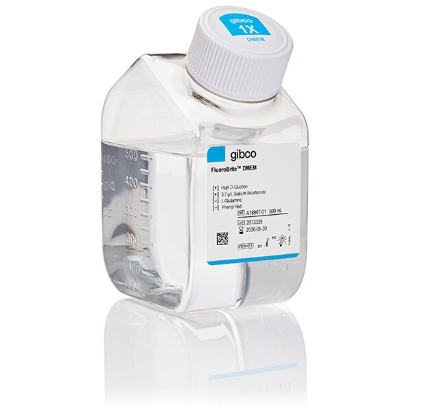
Gibco™
FluoroBrite™ DMEM
Gibco™ FluoroBrite™ DMEMは、バックグラウンド蛍光がPBSと同等であり、標準的なフェノールレッドフリーDMEMよりもバックグラウンド蛍光を90%低下させます。FluoroBrite™ DMEMは、ルーチン細胞培養に10%のウシ胎児血清と4mMのL-グルタミン酸、またはGlutaMAX™サプリメントを添加する場合を想定して、必要な栄養素が最初から含まれており詳細を見る


製品番号(カタログ番号) A1896701
価格(JPY)
6,500
Each
数量:
500 mL
Gibco™ FluoroBrite™ DMEMは、バックグラウンド蛍光がPBSと同等であり、標準的なフェノールレッドフリーDMEMよりもバックグラウンド蛍光を90%低下させます。FluoroBrite™ DMEMは、ルーチン細胞培養に10%のウシ胎児血清と4mMのL-グルタミン酸、またはGlutaMAX™サプリメントを添加する場合を想定して、必要な栄養素が最初から含まれており、フルオロフォアのシグナル/ノイズ比が向上するように設計されています。そのため、最適な細胞の健康を促進する環境で微弱な蛍光事象も可視化できます。その他にも次のような特長があります。
•生細胞イメージング中の蛍光シグナルの改良
• 細胞の健康を維持するために役立つDMEMベース
生細胞蛍光顕微鏡は、根本的に重要で生理学的に関連する生物学的事象を視覚化するために不可欠な技術です。この手法の主要な課題の1つに、弱いフルオロフォアをイメージングする際に必ず細胞の損傷、光退色、または望ましくない細胞の健康変化が伴うことがあります。FluoroBrite™ DMEMは、これらの問題に対処するのに役立ちます。
•生細胞イメージング中の蛍光シグナルの改良
• 細胞の健康を維持するために役立つDMEMベース
生細胞蛍光顕微鏡は、根本的に重要で生理学的に関連する生物学的事象を視覚化するために不可欠な技術です。この手法の主要な課題の1つに、弱いフルオロフォアをイメージングする際に必ず細胞の損傷、光退色、または望ましくない細胞の健康変化が伴うことがあります。FluoroBrite™ DMEMは、これらの問題に対処するのに役立ちます。
研究用にのみ使用できます。診断用には使用いただけません。
仕様
濃度1 X
製造品質cGMP-compliant under the ISO 13485 standard
製品タイプDMEM (Dulbecco's Modified Eagle Medium)
数量500 mL
品質保持期間12 Months From Date of Manufacture
分類Animal Origin-free
形状Liquid
Serum LevelStandard Serum Supplementation
無菌性Sterile-filtered
添加剤ありHigh Glucose
添加剤なしNo Glutamine, No HEPES, No Phenol Red, No Sodium Pyruvate
Unit SizeEach
組成および保存条件
Store in refrigerator (2–8°C). Protect from light.
よくあるご質問(FAQ)
What is the osmolality of Fluorobrite DMEM?
I understand that some media are worse than others for fluorescence imaging. How do I choose?
Should I be concerned about phenol red in my media when labeling my live cells with fluorescent dyes?
引用および参考文献 (21)
引用および参考文献
Abstract
Open source software for quantification of cell migration, protrusions, and fluorescence intensities.
Journal:
PubMed ID:25847537
'Cell migration is frequently accompanied by changes in cell morphology (morphodynamics) on a range of spatial and temporal scales. Despite recent advances in imaging techniques, the application of unbiased computational image analysis methods for morphodynamic quantification is rare. For example, manual analysis using kymographs is still commonplace, often caused by
A BRCA1-interacting lncRNA regulates homologous recombination.
Journal:
PubMed ID:26412854
Long non-coding RNAs (lncRNAs) are important players in diverse biological processes. Upon DNA damage, cells activate a complex signaling cascade referred to as the DNA damage response (DDR). Using a microarray screen, we identify here a novel lncRNA, DDSR1 (DNA damage-sensitive RNA1), which is induced upon DNA damage. DDSR1 induction
Axonal autophagosomes recruit dynein for retrograde transport through fusion with late endosomes.
Journal:
PubMed ID:25940348
Efficient degradation of autophagic vacuoles (AVs) via lysosomes is an important cellular homeostatic process. This is particularly challenging for neurons because mature acidic lysosomes are relatively enriched in the soma. Although dynein-driven retrograde transport of AVs was suggested, a fundamental question remains how autophagosomes generated at distal axons acquire dynein
The Nurr1 Activator 1,1-Bis(3'-Indolyl)-1-(p-Chlorophenyl)Methane Blocks Inflammatory Gene Expression in BV-2 Microglial Cells by Inhibiting Nuclear Factor ?B.
Journal:
PubMed ID:25858541
NR4A family orphan nuclear receptors are an important class of transcription factors for development and homeostasis of dopaminergic neurons that also inhibit expression of inflammatory genes in glial cells. The identification of NR4A2 (Nurr1) as a suppressor of nuclear factor ?B (NF-?B)-related neuroinflammatory genes in microglia and astrocytes suggests that
Posttranslational Modification of HOIP Blocks Toll-Like Receptor 4-Mediated Linear-Ubiquitin-Chain Formation.
Journal:
PubMed ID:26578682
Linear ubiquitination is an atypical posttranslational modification catalyzed by the linear-ubiquitin-chain assembly complex (LUBAC), containing HOIP, HOIL-1L, and Sharpin. LUBAC facilitates NF-?B activation and inflammation upon receptor stimulation by ligating linear ubiquitin chains to critical signaling molecules. Indeed, linear-ubiquitination-dependent signaling is essential to prevent pyogenic bacterial infections that can lead