Search

Reliable 200 kV TEM/STEM imaging with automated workflows
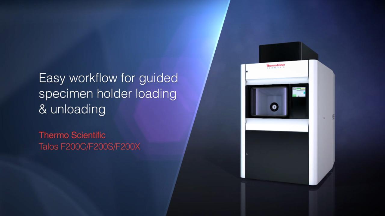
The Thermo Scientific Talos F200C TEM delivers reliable performance with advanced automation in a versatile, multipurpose TEM/STEM platform designed for applications across the life and materials sciences.
Operating at 200 kV with a high-brightness X-FEG source, it provides high resolution and strong penetration capability. The constant-power C-TWIN lens supports enhanced TEM contrast for high-quality imaging. Automated alignments and data collection, combined with streamlined workflows in Thermo Scientific software, help enable faster, more consistent results across a range of user experience levels.
The Talos F200C TEM can be configured with CETA cameras for conventional TEM imaging, integrated STEM and EDX detectors for analytical applications, or the Falcon 4i Direct Electron Detector for cryo-EM workflows. This flexible platform supports a wide range of imaging and diffraction applications.
TEM/STEM components on Talos F200C for high-resolution and analytical imaging
High-Resolution X-FEG
200 kV X-FEG for high resolution and high-penetration depth
C-TWIN lens
Enhanced TEM contrast with the constant-power C-TWIN lens
Fully integrated detector options
Configure with Thermo Scientific Ceta 16M Camera for large field-of-view, fast imaging or Thermo Scientific Falcon 4i Direct Electron Detector for cryo-EM applications.
STEM detectors
Up to four-channel integrated STEM detectors and flexible EDS analysis options
Automated TEM/STEM workflows for reproducibility and ease of use
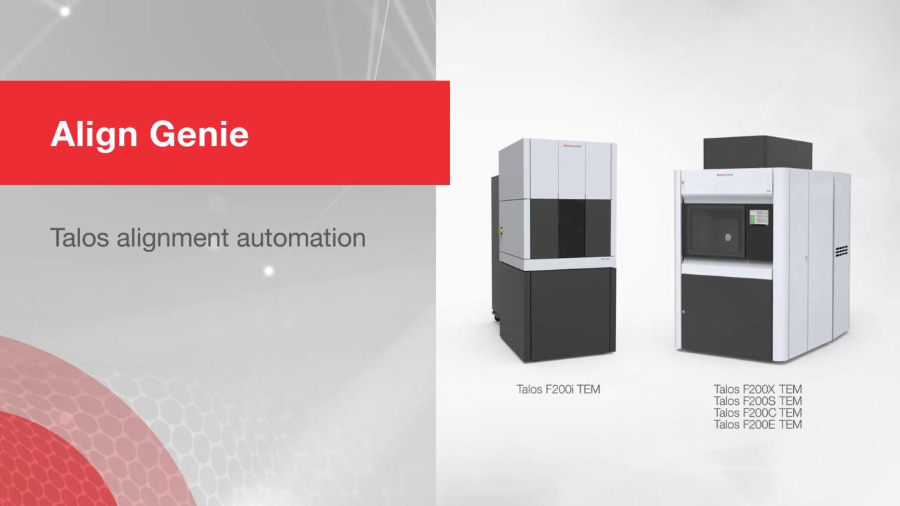
Reproducibility and ease of use with the Talos F200C TEM are supported by automation of daily alignments and image-quality-critical functions.
Commonly used tuning routines are automated within the TEM user interface and provided as standard features. These single-button alignments operate in both TEM and STEM modes and can serve as an alternative to manual alignments.
Key image-quality adjustments are also automated within the application software, offering consistent, single-button control of functions such as autofocus, eucentric height adjustment, and astigmatism correction (autostigmate).
Talos F200C TEM and STEM applications


For Research Use Only. Not for use in diagnostic procedures.