Search

Choose iBright imaging systems based on detection method, imaging requirements, and quantitation needs. Compare western blot imaging capabilities, analysis tools, and system configurations to identify the right solution for imaging, quantitation, and documentation.
Western blot imaging and analysis with iBright imaging systems
Western blot imaging is an important step in protein analysis workflows, enabling researchers to detect, document, and quantify target proteins after electrophoresis and membrane transfer. Whether using chemiluminescent, fluorescent, or colorimetric detection methods, obtaining high-quality images is critical for accurate data interpretation and reproducible results.
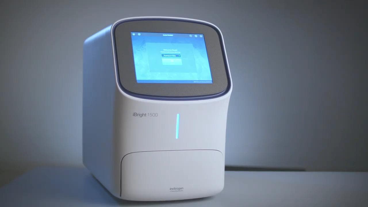
iBright imaging systems combine image acquisition, analysis software, and quantitation tools in a single platform to support western blot imaging, gel documentation, total protein normalization, and protein quantitation.
Whether performing routine western blot analysis or advanced protein expression studies, iBright imaging systems help provide a streamlined solution for imaging, quantitation, and documentation throughout the protein analysis workflow.
Applications and capabilities of iBright imaging systems


Educational resources
Access resources to help optimize western blot detection strategies, troubleshoot experiments, and improve data quality.


Frequently asked questions
A western blot imaging system is an instrument used to visualize, capture, and analyze protein bands on a membrane following western blot detection. Modern imaging systems can support chemiluminescent, fluorescent, and colorimetric detection methods while providing software tools for image analysis and quantitation.
Western blot quantitation is typically performed using densitometry analysis software that measures band intensity. Researchers can compare protein expression levels across samples and normalize results using total protein stains or housekeeping proteins.
Densitometry is a quantitative image analysis method used to measure the intensity of protein bands in a western blot image. The resulting values can be used to evaluate relative protein abundance and compare expression levels between samples.
Smart Exposure technology automatically identifies optimal image acquisition settings to help reduce trial-and-error adjustments and improve imaging consistency across experiments.
For Research Use Only. Not for use in diagnostic procedures.