Search


引用和文献 (8)
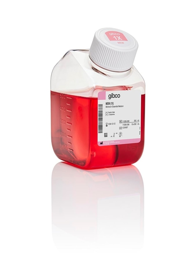
Gibco™
MEM
MEM (Minimum Essential Medium) 是一种较为常用的细胞培养基。MEM 可用于各种悬浮和贴壁的哺乳动物细胞的培养,包括 HeLa、BHK-21、293了解更多信息
Have Questions?
更改视图

| 货号 | 数量 |
|---|---|
| 11095080 | 500 mL |
| 11095098 | 10 x 500 mL |
| 11095072 | 1000 mL |
| 11095114 | 6 x 1000 mL |
货号 11095080
价格(CNY)
385.00
飞享价
Ends: 31-Dec-2026
481.00共减 96.00 (20%)
Each
数量:
500 mL
价格(CNY)
385.00
飞享价
Ends: 31-Dec-2026
481.00共减 96.00 (20%)
Each
MEM (Minimum Essential Medium) 是一种较为常用的细胞培养基。MEM 可用于各种悬浮和贴壁的哺乳动物细胞的培养,包括 HeLa、BHK-21、293、HEP-2、HT-1080、MCF-7、成纤维细胞和原代大鼠星形胶质细胞等。我们针对广泛的细胞培养应用,提供了大量 MEM 改良培养基。使用培养基选择工具查找适合的配方。
这种 MEM 的改良形式如下:
可提供完整配方。
MEM 的使用
MEM 是由 Harry Eagle 在其早期的 Basal Medium Eagle (BME) 配方基础上开发的。此后对 MEM 进行了许多其他改良,包括 Glasgow’s MEM、MEM α、DMEM 以及 Temin’s 改良培养基。MEM 含有 Earle’s 平衡盐用于 CO2 培养箱,或含有 Hanks' 平衡盐用于无 CO2 培养箱。该产品由 Earle’s 平衡盐制成。MEM 不含蛋白质、脂质或生长因子。因此,MEM 需要补充营养成分,通常需要添加 10% 胎牛血清 (FBS)。MEM 使用碳酸氢钠缓冲系统 (2.2 g/L),因此需要 5–10% CO2 的环境来维持生理 pH 值。
cGMP 生产和质量体系
MEM 在位于纽约格兰德岛上符合 cGMP 要求的工厂内生产。该工厂是在FDA登记的医疗器械生产商,且通过ISO 13485标准认证。为确保供应链连续性,我们同时提供由我们的苏格兰工厂生产的等同 MEM 产品 (31095-029)。后者亦是在FDA登记的医疗仪器生产商,且符合ISO 13485标准。
这种 MEM 的改良形式如下:
| 包含 | 不含 |
| • L-谷氨酰胺 | • HEPES: |
| • 酚红 |
可提供完整配方。
MEM 的使用
MEM 是由 Harry Eagle 在其早期的 Basal Medium Eagle (BME) 配方基础上开发的。此后对 MEM 进行了许多其他改良,包括 Glasgow’s MEM、MEM α、DMEM 以及 Temin’s 改良培养基。MEM 含有 Earle’s 平衡盐用于 CO2 培养箱,或含有 Hanks' 平衡盐用于无 CO2 培养箱。该产品由 Earle’s 平衡盐制成。MEM 不含蛋白质、脂质或生长因子。因此,MEM 需要补充营养成分,通常需要添加 10% 胎牛血清 (FBS)。MEM 使用碳酸氢钠缓冲系统 (2.2 g/L),因此需要 5–10% CO2 的环境来维持生理 pH 值。
cGMP 生产和质量体系
MEM 在位于纽约格兰德岛上符合 cGMP 要求的工厂内生产。该工厂是在FDA登记的医疗器械生产商,且通过ISO 13485标准认证。为确保供应链连续性,我们同时提供由我们的苏格兰工厂生产的等同 MEM 产品 (31095-029)。后者亦是在FDA登记的医疗仪器生产商,且符合ISO 13485标准。
仅用于研究和生产用途。不可用于临床诊断或直接用于人类或动物。
规格
细胞系HeLa、BHK-21、293、HEP-2、HT-1080、MCF-7 和成纤维细胞
细胞类型原代大鼠星形胶质细胞
最大浓度1 X
培养环境CO2
适用于(应用)Mammalian Cell Culture
产品规格Bottle
生产质量cGMP-compliant under the ISO 13485 standard
产品线Gibco
产品类型MEM
数量500 mL
有效期自生产之日起 12 个月
运输条件室温
分类非动物源性
培养类型Mammalian Cell Culture
形式液体
血清水平标准血清
无菌无菌过滤
灭菌方法无菌过滤
加有添加剂低糖, 谷氨酰胺, 酚红
不加添加剂不含 HEPES, 不含丙酮酸钠
Unit SizeEach
内容与储存
储存条件:2°C 至 8°C(避光)
运输条件:环境
有效期:自生产之日起 12 个月
运输条件:环境
有效期:自生产之日起 12 个月
常见问题解答 (FAQ)
加入血清后的培养基可以使用多久?
我的培养基是室温条件下运送来的,但注明应保存于冷藏条件下。这会有影响么?
我该如何去除细胞培养基中的支原体污染?
我发现培养物的生长速率变缓。我该如何处理?
我的细胞不能贴附于培养容器。我该如何处理?
引用和文献 (8)
引用和文献
Abstract
Influx of calcium through a redox-sensitive plasma membrane channel in thymocytes causes early necrotic cell death induced by the epipolythiodioxopiperazine toxins.
Journal:J Biol Chem
PubMed ID:12063251
'Gliotoxin, a member of the epipolythiodioxopiperazine (ETP) class of toxins, induces both apoptotic and necrotic cell death in a concentration-dependent manner. Whereas the specific trigger for apoptotic death caused by these toxins is unclear, the reactive disulfide bond in the ETP toxins is required for biological activity. Thus it is
Antagonism between Ena/VASP Proteins and Actin Filament Capping Regulates Fibroblast Motility.
Journal:Cell
PubMed ID:12086607
'Cell motility requires lamellipodial protrusion, a process driven by actin polymerization. Ena/VASP proteins accumulate in protruding lamellipodia and promote the rapid actin-driven motility of the pathogen Listeria. In contrast, Ena/VASP negatively regulate cell translocation. To resolve this paradox, we analyzed the function of Ena/VASP during lamellipodial protrusion. Ena/VASP-deficient lamellipodia protruded
A serum- and antioxidant-free primary culture model of mouse cortical neurons for pharmacological screen and studies of neurotrophic and neuroprotective agents.
Journal:Cell Mol Neurobiol
PubMed ID:12363202
'1. Morphologically developmental properties of fetal mouse cortical neurons in the chemically defined serum- and antioxidant-free culture condition were observed. Also, cellular composition in cultures was identified by immunostaining with anti-NSE and anti-GFAP. 2. Various cell densities ranging from 1 x 10(3) to 1 x 10(6) cells/cm2 were prepared to
Acute hippocampal slice preparation and hippocampal slice cultures.
Journal:Methods Mol Biol
PubMed ID:21815062
'A major advantage of hippocampal slice preparations is that the cytoarchitecture and synaptic circuits of the hippocampus are largely retained. In neurotoxicology research, organotypic hippocampal slices have mostly been used as acute ex vivo preparations for investigating the effects of neurotoxic chemicals on synaptic function. More recently, hippocampal slice cultures,
The Discodermia calyx toxin calyculin a enhances cyclin D1 phosphorylation and degradation, and arrests cell cycle progression in human breast cancer cells.
Journal:Toxins (Basel)
PubMed ID:22069692
'Cyclin D1 is a key regulator of the cell cycle that is over expressed in more than half of breast cancer patients. The levels of cyclin D1 are controlled primarily through post-translational mechanisms and phosphorylation of cyclin D1 at T286 induces its proteasomal degradation. To date, no studies have explored