Search


引用和文献 (14)
Invitrogen™
MycoFluor™ 支原体检测试剂盒
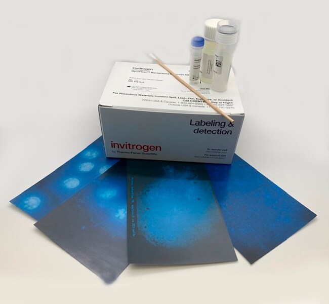
MycoFluor™ 支原体检测试剂盒提供了一种超灵敏、快速和简单的荧光显微镜检测方法,可用于在实验室细胞培养物中目视识别支原体感染。要检测支原体,可以将 MycoFluor™ 荧光试剂直接加入到有细胞或无细胞的培养液中,染色后的样品即可使用荧光显微镜观察。了解更多信息
| 货号 | 数量 |
|---|---|
| M7006 | 100 Tests |
货号 M7006
价格(CNY)
4,716.00
Each
数量:
100 Tests
价格(CNY)
4,716.00
Each
MycoFluor™ 支原体检测试剂盒提供了一种超灵敏、快速和简单的荧光显微镜检测方法,可用于在实验室细胞培养物中目视识别支原体感染。要检测支原体,可以将 MycoFluor™ 荧光试剂直接加入到有细胞或无细胞的培养液中,染色后的样品即可使用荧光显微镜观察。
仅供科研使用。不可用于诊断程序。
规格
检测方法荧光
适用于荧光显微镜
数量100 Tests
运输条件室温
形式液体
产品类型支原体检测
Unit SizeEach
内容与储存
请避光冷藏 (2–8°C) 储存。
常见问题解答 (FAQ)
我该如何去除细胞培养基中的支原体污染?
I suspect mycoplasma is affecting the growth rate of my culture. How can I test for it?
Which fluorescence filter should I use with the MycoFluor Mycoplasma Detection Kit (Cat. No. M7006)?
Can I develop the MycoFluor MycoPlasma Detection Kit (Cat.No. M7006) into a 96-well format for screening with a plate reader?
How can I remove mycoplasma contamination from my cell culture medium?
引用和文献 (14)
引用和文献
Abstract
Characterization of a novel epigenetic effect of ionizing radiation: the death-inducing effect.
Journal:Cancer Res
PubMed ID:12543783
'The detrimental effects associated with exposure to ionizing radiation have long been thought to result from the direct targeting of the nucleus leading to DNA damage; however, the emergence of concepts such as radiation-induced genomic instability and bystander effects have challenged this dogma. After cellular exposure to ionizing radiation, we
Mechanisms of cell death associated with death-inducing factors from genomically unstable cell lines.
Journal:Mutagenesis
PubMed ID:14614192
'We recently described a unique non-targeted effect of ionizing radiation whereby growth medium from two clones of GM10115 cells exhibiting radiation-induced chromosomal instability was cytotoxic to parental GM10115 cells. We termed this the death-inducing effect (DIE). The goal of the present study was to determine how DIE killed cells. Our
Ionizing radiation induces delayed hyperrecombination in Mammalian cells.
Journal:Mol Cell Biol
PubMed ID:15143196
Exposure to ionizing radiation can result in delayed effects that can be detected in the progeny of an irradiated cell multiple generations after the initial exposure. These effects are described under the rubric of radiation-induced genomic instability and encompass multiple genotoxic endpoints. We have developed a green fluorescence protein (GFP)-based
Angiotensin subtype 1 rReceptor (AT1) blockade improves vasorelaxation in heart failure by up-regulation of endothelial nitric-oxide synthase via activation of the AT2 receptor.
Journal:J Pharmacol Exp Ther
PubMed ID:14560036
To determine whether angiotensin receptor blockade decreases vascular tone in heart failure by improving endothelial-dependent vasorelaxation and increasing nitric oxide (NO) bioavailability, we treated infarcted adult male Sprague-Dawley rats with candesartan for 7 days or 8 weeks (10 mg/kg/day in drinking water). Candesartan, at both time points, lowered left ventricular
Combination therapy of BCR-ABL-positive B cell acute lymphoblastic leukemia by tyrosine kinase inhibitor dasatinib and c-JUN N-terminal kinase inhibition.
Journal:J Hematol Oncol
PubMed ID:32552902