Search
What is cryo-electron microscopy?
Cryo-electron microscopy (cryo-EM) is an imaging technique that determines protein structures in high resolution. It is popular for its ability to visualize large, macromolecular complexes including membrane proteins, which are important targets for drug development and understanding biological processes.
Using only small amounts of sample, cryo-EM can collect atomic-resolution images even for complexes that are difficult to prepare as well as those that do not crystallize readily. Ongoing technological advances like direct electron detectors and powerful software have made cryo-EM an indispensable tool for exploring the structure and function of biological molecules, especially those previously considered intractable.
Cryo-EM techniques: Imaging single particles in vitro and cellular structures in situ
Cryo-electron microscopy (cryo-EM) encompasses several methods for visualizing biological structures at high resolution, including two particularly powerful approaches: single particle analysis and cryo-electron tomography (cryo-ET).
Single particle analysis is used to determine the 3D structures of purified macromolecules by imaging and then computationally averaging thousands to millions of individual particles frozen in vitreous ice. This technique is ideal for studying isolated proteins, complexes, and viruses and for achieving the highest resolutions possible with cryo-EM.
Cryo-electron tomography (cryo-ET) captures 3D snapshots of larger volumes of ultra-thin cell and tissue sections that have been frozen and then thinned using a cryo-FIB. The frozen lamella is tilted in the cryo-TEM, which takes a series of images that are reconstructed into a tomogram, revealing cellular macromolecules, structures, and organelles in context within their native environments.


What is cryo-EM used for?
Cryo-EM opens transformative opportunities for researchers to study biological macromolecules. It provides structural insights that add a crucial layer of understanding by revealing molecular form, thereby clarifying how shape dictates function.
Cryo-EM can uncover the precise mechanisms of proteins. For example, it helps researchers understand DNA replication and transcription, and it supports gene editing technologies like CRISPR.
The structure of the 70S ribosome shown here, captured using single particle analysis, provides deep insights into how proteins are made in prokaryotes like bacteria and offers targets for antibiotic development.

Cryo-EM illuminates the mechanisms of antigen recognition by antibodies, informing vaccine design and immunotherapies.
Cryo-EM-based structural biology helped solve this structure of respiratory syncytial virus (RSV) nucleocapsid-like assemblies, providing information on the virus architecture and contributing to the design of effective RSV vaccine candidates.

Analyzing synapses with cryo-ET can reveal the mechanism of neurotransmission through ion channel structures.
This cryo-ET image of an excitatory synapse of a hippocampal neuron clearly depicts post-synaptic structure-like PSD filaments and glutamate receptors as well as pre-synaptic structures such as vesicles and adhesion proteins. Courtesy of Guoqiang Bi and Hong Zhou of University of California, Los Angeles.

Cryo-EM can help researchers understand the diverse structural arrangements of amyloid filaments associated with a wide array of neurodegenerative diseases.
The image here depicts Alzheimer’s disease associated TMEM106B filaments as described by Chang et al. (PDB ID: 7U11). They showed that a previously unsolved amyloid fibril is composed of TMEM106B, a transmembrane lysosomal/endosomal protein.

Cryo-EM delivers structural insights into membrane proteins, such as transporters and G protein-coupled receptors, enabling rational drug design.
The GABAA receptor shown here is a key target in structure-based drug design targeting neurological disorders such as anxiety, epilepsy, insomnia, and depression.

Cryo-EM makes it possible to map viral entry mechanisms, such as those of HIV and SARS-CoV-2, paving the way for antiviral drug development.
Cryo-EM has contributed to our understanding of SARS-CoV-2 since the beginning of the COVID-19 pandemic. Shown here is a 3D volume rendering of SARS-CoV-2 virion budding and assembly at the ERGIC membrane captured using cryo-ET.
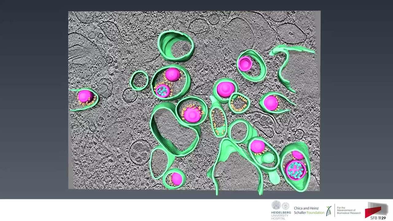
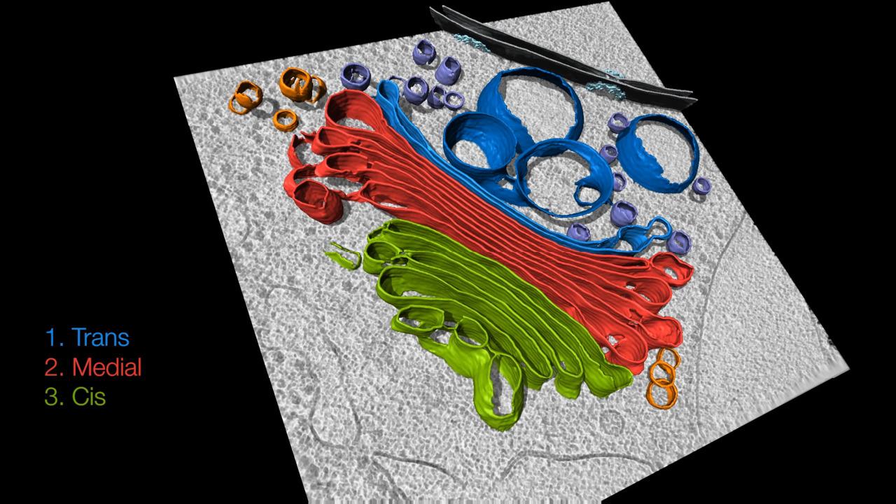
Cryo-EM reveals the architecture and spatial organization of macromolecular complexes within cells, helping researchers understand processes like intracellular transport, organelle dynamics, and cytoskeletal remodeling in their native context.
Get cryo-EM access and training at supporting centers
Access to a cryo-transmission electron microscope (cryo-TEM) can be a significant barrier for many researchers given the cost and technical expertise required to operate these advanced instruments. However, national research infrastructures in both Europe and North America are working to remove these barriers by providing open access to state-of-the-art cryo-EM facilities.
These initiatives ensure that scientists without local cryo-EM capabilities can obtain training and apply the technique to their research, fostering broader innovation and collaboration across the structural biology community.
Cryo-EM centers in Europe


In Europe, Instruct‑ERIC offers centralized access to high-end cryo-EM technologies and expert support across its member countries. It also provides hands-on training and funded access for eligible researchers.
Cryo-EM centers in North America


In North America, centers provide access to cutting-edge instruments, comprehensive training programs, and data processing support—regardless of institutional affiliation.
Cryo-EM instruments to build or expand your facility
When building or expanding a cryo-EM facility, choosing the right instrumentation is key to ensuring user accessibility and long-term success. Thermo Scientific transmission electron microscopes are specifically designed with new users in mind, featuring advanced automation, intuitive user interfaces, and streamlined workflows that significantly lower the learning curve.
From sample loading to data acquisition, Thermo Scientific TEMs optimize every step to reduce complexity and maximize reproducibility, ultimately delivering results faster—even for users with limited prior experience. These instruments help facilities support a broader user base, accelerate training, and deliver high-quality data with greater efficiency, making them an outstanding foundation for a scalable and user-friendly cryo-EM program.


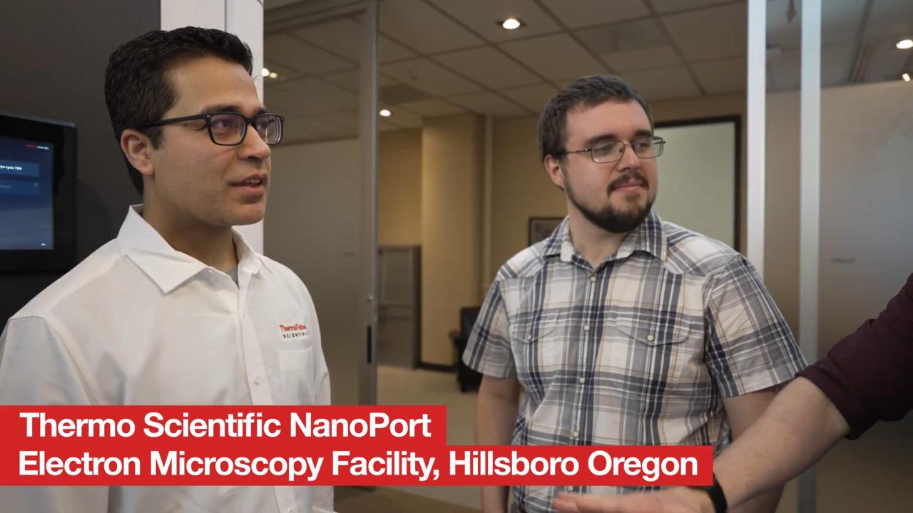
Get hands-on experience with Thermo Scientific TEMs at our NanoPort Facilities
Thermo Fisher Scientific’s NanoPort Facilities are global hubs designed to provide hands‑on access to the latest electron microscopy technologies. Whether you're new to electron microscopy or seeking to test advanced instrumentation with your own samples, these centers offer expert-led workshops, instrument demonstrations, and training to help you evaluate and adopt the right tools. By visiting, you can see how instruments perform in realistic settings, collaborate with specialists, and stay on the cutting edge of microscopy advances. Visit us in North America, Europe, and Asia.

Funding electron microscopy lab and facility equipment
Thermo Fisher’s Funding Support Center offers resources to help you secure funding for acquiring, upgrading, and sustaining electron microscopy equipment. Whether you’re new to electron microscopy or experienced, we offer consultative support to help you navigate grants, internal funding, and other financial sources, including flexible financing options.
To help strengthen your proposal, our team can help you collect preliminary datasets at our NanoPort Facilities and provides insight into current technology trends and applications.

Get support from our cryo-EM experts
Whether you are just starting to learn about cryo-EM or are in the process of adopting the technology, our experts are here to help with everything from initial planning to data collection and interpretation. Reach out to us today to learn more about how cryo-EM techniques and instruments can help advance your research.
For Research Use Only. Not for use in diagnostic procedures.