Search
How cryo-EM enables antibody discovery and epitope mapping
Understanding how antibodies interact with their target antigens is critical for successful antibody discovery and development. Structural characterization of antibody–antigen interactions enables more informed candidate selection, improved differentiation, and reduced risk across development workflows.
As of 2026, more than 160 monoclonal antibody therapies have been approved globally, highlighting the growing importance of robust and efficient antibody discovery strategies.
Cryo-electron microscopy (cryo-EM) enables high-resolution structural analysis of antibody–antigen complexes in near-native conditions, including systems that are difficult to study using traditional techniques such as X-ray crystallography or HDX-MS. By directly visualizing binding interfaces without the need for crystallization or labeling, cryo-EM reveals how antibodies recognize their targets and provides detailed insight into the mechanism of action.
Structural insight from cryo-EM can help:
- Accelerate antibody candidate selection
- Clarify binding specificity and interaction geometry
- Differentiate between competing antibody candidates
- Inform downstream development decisions

These capabilities make cryo-EM a powerful approach for epitope mapping, a key step in antibody discovery workflows.
Epitope mapping with cryo-EM
Epitope mapping is a critical component of antibody discovery, enabling precise identification of how and where antibodies bind to their target antigens. This information underpins understanding of specificity, selectivity, and the mechanism of action, and it can contribute to candidate differentiation and intellectual property (IP) strategies.
Epitopes can be broadly classified into two types: linear epitopes and conformational epitopes, the latter of which are commonly targeted by therapeutic antibodies.
Cryo-EM enables structural epitope mapping at near-atomic resolution by directly visualizing epitope–paratope interactions. This approach allows detailed characterization of binding interfaces and direct comparison of binding modes across antibody candidates.
 Cryo-EM enables visualization of epitope–paratope interactions at the amino acid level, supporting detailed understanding of antibody binding and the mechanism of action.
Cryo-EM offers several advantages for antibody epitope mapping:
- High-resolution mapping of epitope–paratope interactions at the amino acid level
- Direct comparison of binding modes across multiple antibody candidates
- Characterization of heterogeneous or flexible antigen targets
- Simultaneous analysis of multiple antibody–antigen complexes in a single experiment
- No requirement for crystallization, labeling, or immobilization, avoiding common structural artifacts
By overcoming key limitations of traditional structural biology methods, cryo-EM expands the range of antibody–antigen systems that can be structurally characterized.
Epitope mapping for therapeutic antibody development using cryo-EM
Structural epitope mapping using cryo-EM supports the development of therapeutic antibodies by enabling detailed characterization of binding behavior and the mechanism of action.
These insights can support:
- Design and optimization of antibody formats such as bispecific antibodies and antibody-drug conjugates (ADCs)
- Structural comparison of candidate binding modes for differentiation
- Selection of lead candidates based on binding properties
Importantly, amino acid–level epitope definition can strengthen intellectual property (IP) strategies by demonstrating binding specificity, novelty, and non-obviousness.
 Example of cryo-EM-based analysis of multiple antibody–antigen complexes, supporting epitope mapping and comparison of candidate binding interactions within a single experiment.
Epitope mapping to develop antibodies for use as a tool or reagent
Due to their remarkable selectivity, antibodies are routinely used to develop highly specific diagnostic tests, and affinity resins are used in bioprocessing and downstream purification. More recently, antibody-derived fragments are also being used as tools to study other therapeutically relevant target proteins such as GPCRs and ion channels. Some of these tools are nanobody, megabody, Legobody, and NabFab.

Cryo-EM structure of the PepT2 solute transporter stabilized by a nanobody, enabling structural characterization of a membrane protein complex. Image adapted from Parker et al, 2021.

Cryo-EM structure of the SARS-CoV-2 spike protein receptor-binding domain (RBD) in complex with an engineered antibody fragment (Legobody), illustrating the use of antibody-based reagents to enable structural analysis. Image adapted from Wu and Rapoport, 2021.
AI-driven antibody discovery
Artificial intelligence (AI) and machine learning (ML) approaches are increasingly used to design and optimize antibody candidates by predicting structures and binding interactions.
In these workflows, experimental validation of predicted binding modes is essential.
Cryo-EM provides high-resolution structural data that enables:
- Validation of predicted antibody–antigen interactions
- Assessment of structural accuracy
- Refinement of computational models
By combining AI-based design with experimental structural data, cryo-EM supports iterative, structure-guided antibody discovery workflows.


Cryo-EM structure of a de novo designed VHH (nanobody) bound to influenza hemagglutinin, supporting validation of predicted binding modes, structural accuracy, and CDR loop conformation. Image adapted from Bennett et al., 2026.
Antibody discovery workflow
Cryo-EM provides structural insight that can be applied across multiple stages of the antibody discovery and development workflow. From early research to clinical evaluation, it enables detailed characterization of antibody–antigen interactions, supporting more informed decisions at each stage.
By integrating structural data throughout the workflow, cryo-EM helps accelerate candidate progression, improve differentiation, and reduce development risk.
Notably, of antibody therapeutics entering Phase 1 clinical trials, only approximately 22% ultimately receive regulatory approval—highlighting the importance of early, high-quality structural insight.
 Cryo-EM can provide structural insight across multiple stages of the antibody discovery workflow, supporting characterization, optimization, and evaluation of candidate molecules.
- Hit to lead
- Lead optimization
- Candidate selection
- Clinical trials
Hit to lead in therapeutic antibody development
During early-stage discovery, large numbers of candidate antibodies are generated and screened. Structural characterization at this stage helps prioritize the most promising candidates.
Cryo-EM enables:
- Structural characterization of antibody–antigen binding interfaces
- High-resolution mapping of epitopes and paratopes
- Direct comparison of candidate binding modes
- Early identification of candidates with favorable binding properties
 Antibodies can recognize linear or conformational epitopes, which can be characterized using structural methods such as cryo-EM.
Lead optimization
In the lead optimization phase, antibody candidates are refined to improve affinity, specificity, and stability.
Cryo-EM supports structure-guided optimization by providing:
- Structural insight to guide rational antibody engineering
- Evaluation of the impact of sequence modifications
- Input for computational modeling and molecular dynamics simulations
- Experimental validation of optimized designs
Candidate selection for therapeutic antibody development
Selection of candidates for clinical development requires a detailed understanding of binding behavior and the mechanism of action.
Cryo-EM enables:
- High-resolution epitope mapping to define the mechanism of action
- Structural comparison of candidates for differentiation
- Identification of unique binding modes supporting IP strategies
- Generation of structural data packages supporting regulatory submissions
Clinical trials
During preclinical and clinical development, understanding immune response and safety profiles is essential.
Cryo-EM can support:
- Structural characterization of immune responses to therapeutic antibodies
- Analysis of antibody–antigen and immune complex interactions
- Insight into immunogenicity and the biological mechanism of action
A continuous structural data trail from discovery through clinical stages enhances scientific rigor and supports regulatory expectations.
Cryo-EM instruments for antibody discovery and epitope mapping
The Thermo Scientific cryo-electron microscopes featured here are designed to support antibody discovery, epitope mapping, and structural biology workflows.
These instruments enable high-resolution structural characterization of antibody–antigen interactions, supporting critical decisions across drug discovery—from early screening to lead optimization and candidate selection.


Resources
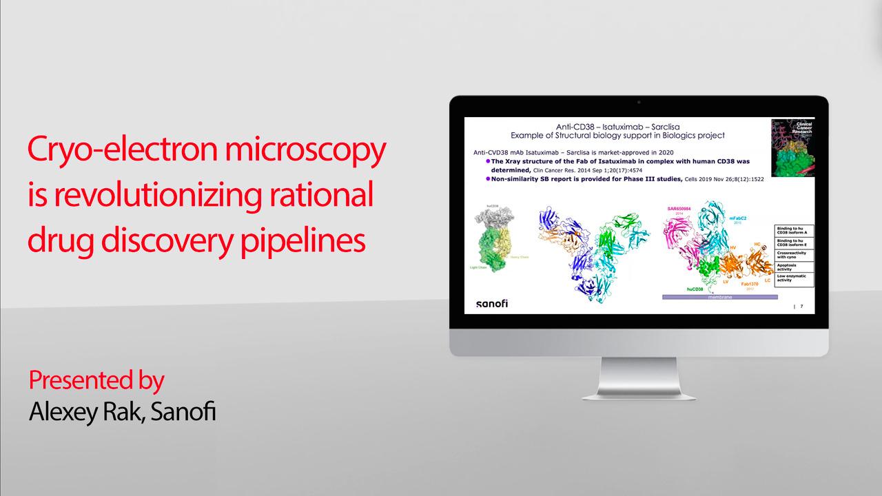
Webinar: Cryo-electron microscopy is revolutionizing rational drug discovery pipelines
Rational design leveraging routine, high-resolution protein structure determination is driving the discovery and development of diverse biologic and small molecule therapies. Cryo-EM delivers rapid epitope mapping on the atomic scale for antibody therapeutics and immune response profiling, supports elucidation of the mechanism of action, and enables more therapeutic targets than ever before for structure-based drug design. Whether you’re modulating binding affinities or optimizing drug stability, all of these questions can be answered in just one day of data collection.
Watch the webinar to hear experts from Sanofi discuss:
- An overview of how to quickly go from protein to structure
- Key examples of multi-specific drugs and structural insights of a CEACAM5-targeting antibody drug conjugate (ADC)
- How the adoption of cryo-EM has significantly increased the number of the targets that can be identified for clinical trials in both biopharma and biotech


Frequently asked questions
Epitope mapping identifies the specific region (epitope) on an antigen that is recognized by an antibody. This information is critical for understanding the mechanism of action, guiding antibody engineering, and supporting candidate selection. Structural characterization of antibody–antigen interactions can also contribute to intellectual property claims and differentiation of therapeutic candidates.
Epitopes are typically classified into linear epitopes (continuous amino acid sequences) and conformational epitopes (discontinuous residues brought together by protein folding). Most therapeutic antibodies recognize conformational epitopes, which require structural methods for accurate characterization.
Cryo-EM enables visualization of antibody–antigen complexes at near-atomic resolution under near-native conditions. It allows:
- Direct observation of binding interfaces
- Mapping of both epitope and paratope
- Analysis of flexible or complex targets that may be challenging for other structural methods
This structural information helps interpret binding interactions and supports downstream optimization.
Cryo-EM can be applied across multiple stages:
- Hit-to-lead: identify binding modes and epitope location
- Lead optimization: guide structure-based engineering
- Candidate selection: compare binding mechanisms and select leads
- Preclinical and clinical: assess immunogenicity and the mechanism of action
Early access to structural data can inform decision-making throughout the workflow.
Structural information enables:
- Identification of key binding residues
- Rational modification of antibody regions (e.g., CDRs)
- Assessment of steric clashes or binding limitations
- Improved understanding of specificity and affinity
This supports more informed and targeted engineering compared to sequence-only approaches.
Yes. Cryo-EM provides experimental structural data that can:
- Confirm predicted antibody–antigen interactions
- Validate binding conformations
- Improve training datasets for AI and ML models
Experimental validation remains important to confirm predicted designs and assess their functional relevance.
By resolving antibody–antigen complexes, cryo-EM can:
- Identify binding sites and interaction geometry
- Reveal inhibition mechanisms (e.g., steric blocking, receptor clustering)
- Provide insight into functional effects at the molecular level
This structural context supports interpretation of biological activity.
Cryo-EM offers:
- Structural visualization without labeling or crystallization
- Applicability to large, flexible, or heterogeneous complexes
- Ability to resolve multiple binding modes or stoichiometries
However, it is typically used alongside complementary biochemical and biophysical methods.
In optimized workflows, cryo-EM can support analysis of multiple antibody–antigen complexes within a single experiment. This can enable efficient evaluation of candidate interactions and support comparative analysis across antibody panels.
Yes. Structural insights from cryo-EM can inform the design of:
- Monoclonal antibodies
- Bispecific antibodies
- Antibody-drug conjugates (ADCs)
- Antibody fragments (e.g., nanobodies)
Understanding spatial binding constraints is particularly important for multi-specific formats.
While powerful, cryo-EM has considerations:
- Requires specialized instrumentation and expertise
- Resolution may vary depending on sample quality and complexity
- Data interpretation depends on accurate modeling
It is most effective when integrated with complementary analytical and computational approaches.
No. Cryo-EM is complementary to techniques such as X-ray crystallography, NMR, and HDX-MS. Each method provides different types of information, and combined approaches are often used for comprehensive characterization.
Detailed structural characterization can:
- Demonstrate binding specificity at the amino acid level
- Support claims of novelty and non-obviousness
- Provide evidence of the mechanism of action
Such data may strengthen regulatory submissions and patent applications.
For Research Use Only. Not for use in diagnostic procedures.